. 2012;7(12):e52330.
doi: 10.1371/journal.pone.0052330. Epub 2012 Dec 21.

Modelling co-infection of the cystic fibrosis lung by Pseudomonas aeruginosa and Burkholderia cenocepacia reveals influences on biofilm formation and host response

Affiliations

Modelling co-infection of the cystic fibrosis lung by Pseudomonas aeruginosa and Burkholderia cenocepacia reveals influences on biofilm formation and host response

Alessandra Bragonzi et al. PLoS One. 2012.

Abstract

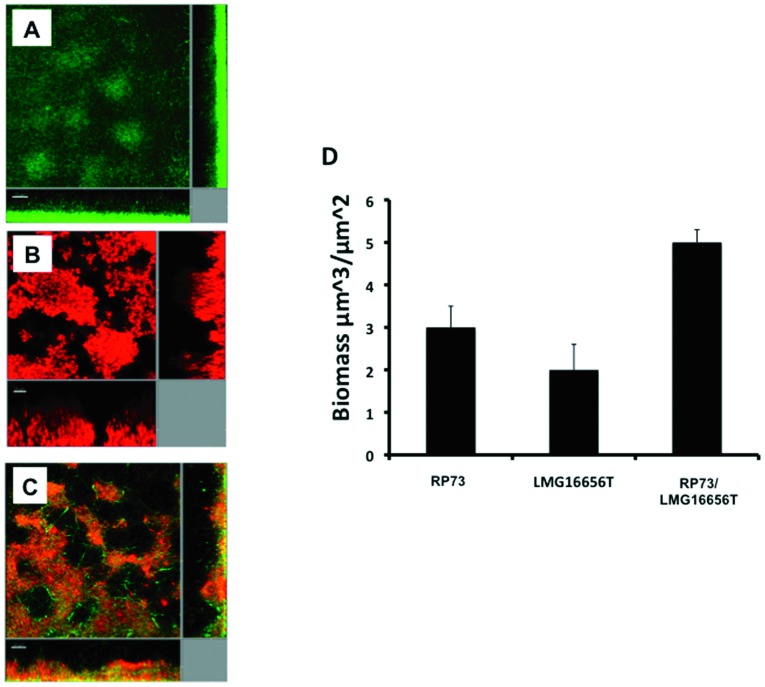
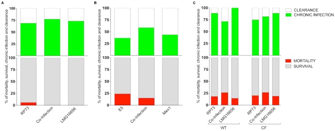
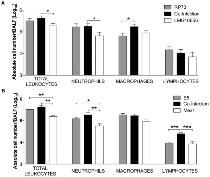
The Gram-negative bacteria Pseudomonas aeruginosa and Burkholderia cenocepacia are opportunistic human pathogens that are responsible for severe nosocomial infections in immunocompromised patients and those suffering from cystic fibrosis (CF). These two bacteria have been shown to form biofilms in the airways of CF patients that make such infections more difficult to treat. Only recently have scientists begun to appreciate the complicated interplay between microorganisms during polymicrobial infection of the CF airway and the implications they may have for disease prognosis and response to therapy.To gain insight into the possible role that interaction between strains of P. aeruginosa and B. cenocepacia may play during infection, we characterised co-inoculations of in vivo and in vitro infection models. Co-inoculations were examined in an in vitro biofilm model and in a murine model of chronic infection. Assessment of biofilm formation showed that B. cenocepacia positively influenced P. aeruginosa biofilm development by increasing biomass. Interestingly, co-infection experiments in the mouse model revealed that P. aeruginosa did not change its ability to establish chronic infection in the presence of B. cenocepacia but co-infection did appear to increase host inflammatory response.Taken together, these results indicate that the co-infection of P. aeruginosa and B. cenocepacia leads to increased biofilm formation and increased host inflammatory response in the mouse model of chronic infection. These observations suggest that alteration of bacterial behavior due to interspecies interactions may be important for disease progression and persistent infection.


Conflict of interest statement

Competing Interests: The authors have declared that no competing interests exist.

Figures

Figure 1
Figure 1. Single and dual species batch growth curves and competitive index values.
The two species were individually cultured or co-cultured at a 1∶1 ratio and grown for 24 h in NB medium at 37°C with vigorous aeration. Colony-forming unit counts (CFU) were determined at 0, 2, 4, 6, 8 and 24 h of bacterial growth. The results are the mean of Log (CFU ml−1) values of three separated assays. Key: (A) Growth of clinical P. aeruginosa RP73 and B. cenocepacia LMG16656 strains in single and dual cultures; (B) Competitive index (CI) and relative increase ratio (RIR) generated from single and dual cultures of clinical P. aeruginosa RP73 and B. cenocepacia LMG16656 strains; (C) Growth of environmental P. aeruginosa E5 and B. cenocepacia Mex1 strains in single and dual cultures; (D) Competitive index (CI) and relative increase ratio (RIR) generated from single and dual cultures of environmental P. aeruginosa E5 and B. cenocepacia Mex1 strains. CI and RIR were calculated as described in Materials and Methods . Each value represents the mean of RIR and CI values from three separate assays, and the bars indicate standard deviations. * = P<0.05, ** = P<0.01 in the Student's t test.
Figure 2
Figure 2. Biofilm formation by P. aeruginosa and B. cenocepacia strains in single and dual cultures.
Bacteria were grown overnight in 96-well polyvinyl chloride flat-bottomed microtiter plates in NB medium at 37°C either individually cultured or co-cultured at a 1∶1 ratio or when individually cultured supplemented with sterile concentrated supernatant of the second organism at a final concentration of 1×. Biofilm biomass was quantified by staining with crystal violet and absorbance measurements at OD 595. The values are means of three separated assays, and the bars indicate standard deviation. * = P<0.05, ** = P<0.01, *** = P<0.001 in Student's t test. S = supernatant.
Figure 3
Figure 3. P. aeruginosa and B. cenocepacia planktonic and sessile cells in single and dual cultures.
Bacteria were grown overnight in 96-well polyvinyl chloride flat-bottomed microtiter plates in NB medium at 37°C either individually cultured or co-cultured at a 1∶1 ratio. CFU counts were determined at 24 h of bacterial growth in both planktonic and sessile fraction. Key: (A, left) Sessile cells of clinical pair (P. aeruginosa RP73 and B. cenocepacia LMG16656) in single and dual cultures; (A, right) Sessile cells of environmental pair (P. aeruginosa E5 and B. cenocepacia Mex1) in single and dual cultures; (B, left) Planktonic cells of clinical pair (P. aeruginosa RP73 and B. cenocepacia LMG16656) in single and dual cultures; (B, right) Planktonic cells of environmental pair (P. aeruginosa E5 and B. cenocepacia Mex1) in single and dual cultures; (C) CI and RIR mean values of sessile growth of P. aeruginosa versus B. cenocepacia (RP73 versus LMG16656, E5 versus Mex1); (D) CI and RIR of planktonic growth of P. aeruginosa versus B. cenocepacia. Each value represents the mean of RIR and CI values from three separate assays, and the bars indicate standard deviations. * = P<0.05, ** = P<0.01, *** = P<0.001 in Student's t test.
Figure 4
Figure 4. Biofilm architecture in P. aeruginosa is influenced by B. cenocepacia.
Images are of 4-day-old biofilms in flow cells in FABL medium. Key: (A) P. aeruginosa RP73; (B) B. cenocepacia LMG16656;(C) mixed culture of P. aeruginosa RP73 and B. cenocepacia LMG16656; (D) Quantification of biomass as determined using COMSTAT to estimate the percentage of P. aeruginosa cells as a function of the total biomass. For these experiments, P. aeruginosa was tagged with mini-Tn7gfp. B. cenocepacia was visualized with Syto62, as described in Materials and Methods . Scale bars = 20 µm. Images shown are representative of 12 images from three independent experiments.
Figure 5
Figure 5. Virulence of P. aeruginosa and B. cenocepacia strains alone or in co-infection in mice.
C57Bl/6 mice (A and B), Cftrtm1UNCTgN(FABPCFTR) (CF) and their congenic wt mice (C) were infected with P. aeruginosa and/or B. cenocepacia strains. Mortality induced by bacteremia (red) and survival (grey) were evaluated on challenged mice. Clearance (white) and capacity to establish chronic airways infection (green) after 13 days from challenge were determined on surviving mice infected with P. aeruginosa and B. cenocepacia strains alone or with pairs of clinical (A and C) or environmental (B) strains. The data are pooled from two to three independent experiments. Mortality and chronic infection are reported as median values. B6.129P2-Cftrtm1UNCTgN(FABPCFTR) Cftr +/+ and B6.129P2-Cftrtm1UNCTgN(FABPCFTR)CftrS489X/S489X mice co-infected with RP73-LMG16656 developed a higher rate of mortality when compared with C57BL/6NCrlBR mice (P<0.05; see Table S1).
Figure 6
Figure 6. Total and differential cell counts in BAL fluid after 13 days of infection.
The number of total leukocytes and in particular of neutrophils, monocytes and lymphocytes recruited in the airways were analyzed in BAL fluid (BALF) after 13 days of chronic lung infection with pairs of clinical (A) or environmental strains (B)). Values represent the mean ± SEM. The data are pooled from two or three independent experiments. Statistical significance by two tailed Student's t-test is indicated: * P<0.05, ** P<0.01.

Similar articles

Cited by

References

    1. Sibley CD, Rabin H, Surette MG (2006) Cystic fibrosis: a polymicrobial infectious disease. Future Microbiol 1: 53–61. - PubMed
    1. Rajan S, Saiman L (2002) Pulmonary infections in patients with cystic fibrosis. Semin Respir Infect 17: 47–56. - PubMed
    1. Lipuma JJ (2010) The changing microbial epidemiology in cystic fibrosis. Clin Microbiol Rev 23: 299–323. - PMC - PubMed
    1. Gilligan PH (1991) Microbiology of airway disease in patients with cystic fibrosis. Clin Microbiol Rev 4: 35–51. - PMC - PubMed
    1. Govan JR, Deretic V (1996) Microbial pathogenesis in cystic fibrosis: mucoid Pseudomonas aeruginosa and Burkholderia cepacia. Microbiol Rev 60: 539–574. - PMC - PubMed

Publication types