. 2012;7(12):e52465.
doi: 10.1371/journal.pone.0052465. Epub 2012 Dec 20.

Gastric cancer exosomes trigger differentiation of umbilical cord derived mesenchymal stem cells to carcinoma-associated fibroblasts through TGF-β/Smad pathway

Affiliations

Gastric cancer exosomes trigger differentiation of umbilical cord derived mesenchymal stem cells to carcinoma-associated fibroblasts through TGF-β/Smad pathway

Jianmei Gu et al. PLoS One. 2012.

Abstract

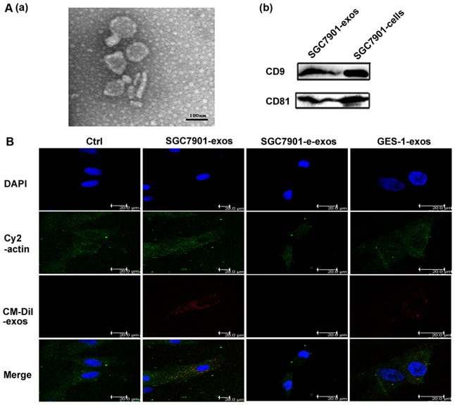
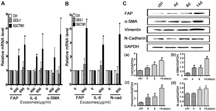
Background: Mesenchymal stem cells (MSCs) promote tumor growth by differentiating into carcinoma-associated fibroblasts (CAFs) and composing the tumor microenvironment. However, the mechanisms responsible for the transition of MSCs to CAFs are not well understood. Exosomes regulate cellular activities by mediating cell-cell communication. In this study, we aimed to investigate whether cancer cell-derived exosomes were involved in regulating the differentiation of human umbilical cord-derived MSCs (hucMSCs) to CAFs.

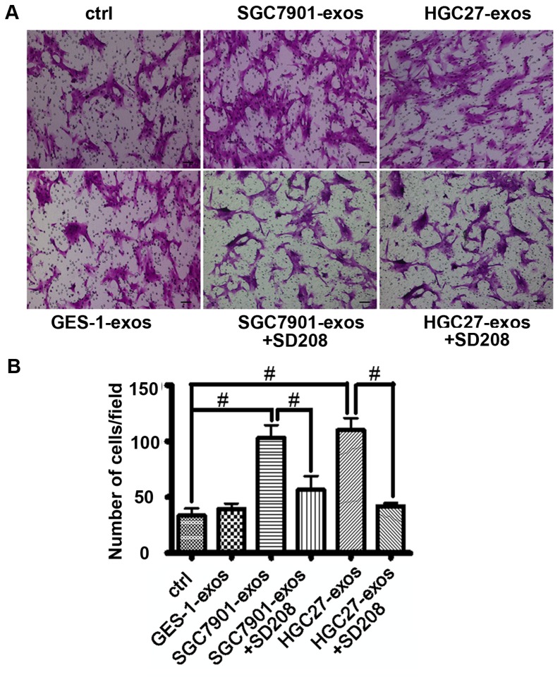
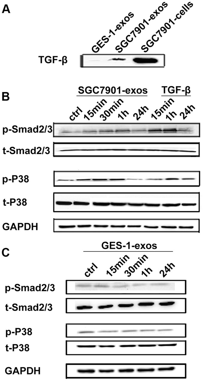
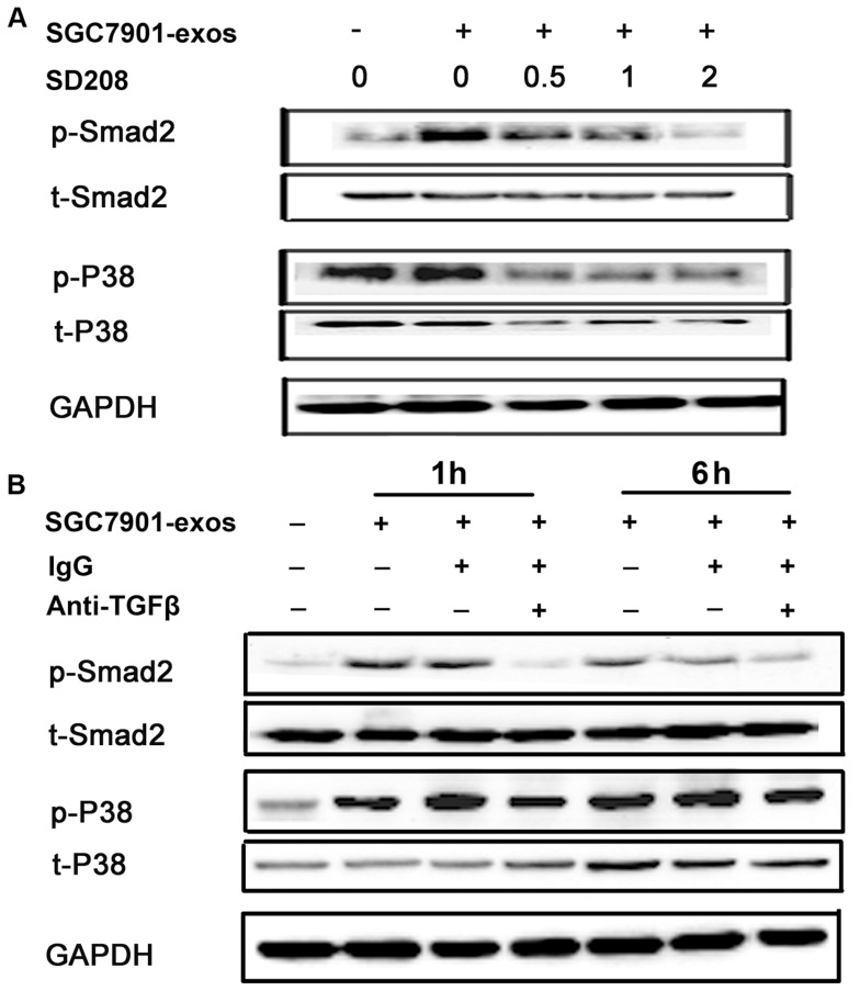
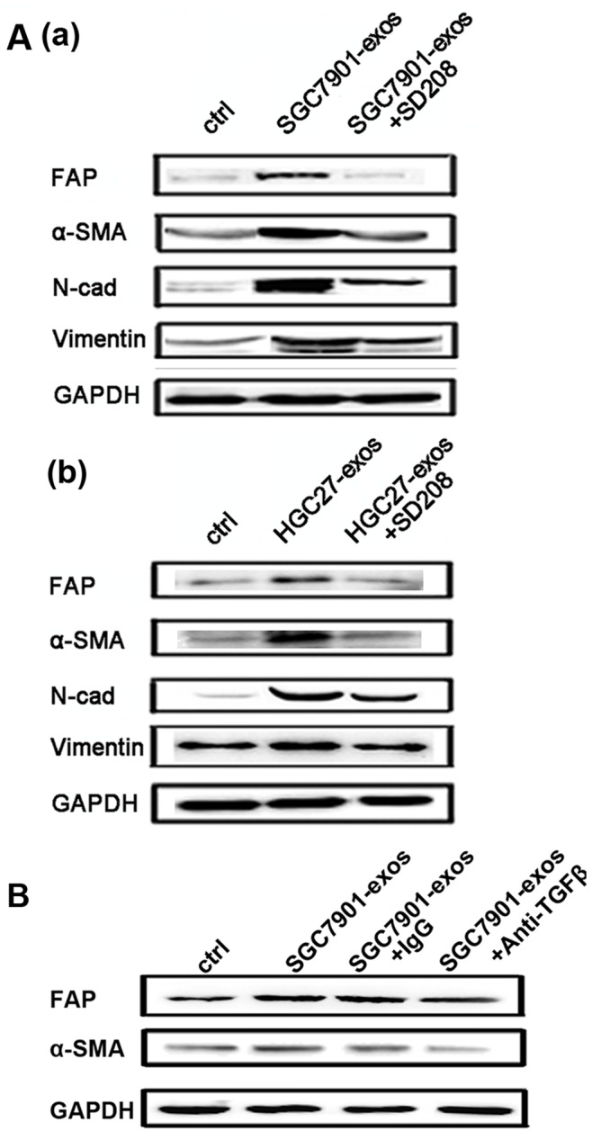
Methodology/principal findings: We first showed that gastric cancer cell-derived exosomes induced the expression of CAF markers in hucMSCs. We then demonstrated that gastric cancer cell-derived exosomes stimulated the phosphorylation of Smad-2 in hucMSCs. We further confirmed that TGF-β receptor 1 kinase inhibitor attenuated Smad-2 phosphorylation and CAF marker expression in hucMSCs after exposure to gastric cancer cell-derived exosomes.

Conclusion/significance: Our results suggest that gastric cancer cells triggered the differentiation of hucMSCs to CAFs by exosomes-mediated TGF-β transfer and TGF-β/Smad pathway activation, which may represent a novel mechanism for MSCs to CAFs transition in cancer.


Conflict of interest statement

Competing Interests: The authors have declared that no competing interests exist.

Figures

Figure 1
Figure 1. Exosomes characterization and MSC internalization.
A. The morphology of exosomes and exosomal marker expression. (a) Morphologic analysis of gastric cancer derived exosomes. Scale bar = 100 nm. (b) CD9 and CD81 expression in exosomes. B. Exosomes were uptaken by hucMSCs. Exosomes labeled with CM-Dil (red) at 37°C for 1 h were added into hucMSCs and incubated for 4 h. Effluent from a filtered suspension of CM-Dil-labeled exosomes (control) and the CM-Dil-labeled withdrawn exosomes fraction (e-exos) were used as controls. Cells were fixed and stained for cytoplasm (cy2-actin, green) and nuclei (DAPI, blue). Scale bar = 20 µm.
Figure 2
Figure 2. Gastric cancer cell derived exosomes induces the expression of CAF markers in hucMSCs.
A.Quantitative analyses of the FAP, IL-6 and α-SMA mRNA expression in hucMSCs. HucMSCs were treated with various concentrations of SGC7901 exosomes or GES-1 exosomes for 36 hours. B. Quantitative analyses of the FAP, IL-6 and N-Cadherin expression in hucMSCs. HucMSCs were treated with various concentrations of SGC7901 exosomes or GES-1 exosomes for 14 days. C. Western blotting analyses of the FAP, α-SMA, N-Cadherin and Vimentin protein expression in hucMSCs treated with SGC7901 exosomes (800 µg/ml) for different times. (a–d) Density analysis of Western blotting bands. *P<0.05 and # P<0.01, compared to the relative control group (n = 3).
Figure 3
Figure 3. Gastric cancer cell derived exosomes promote hucMSCs migration.
A. HucMSCs were treated with gastric cancer cell or normal gastric epithelial cell derived exosomes (800 µg/mL) in the presence or absence of SD208 (2 µM) for 6 h. Transwell migration assay was performed to analyze the migratory ability of the cells. B. The number of migrated cells above was evaluated. These experiments were repeated for three times. *P<0.05 and # P<0.01, n = 3. Scale bar = 50 µm.
Figure 4
Figure 4. Gastric cancer cell derived exosomes induce Smad2/3 and p38 phosphorylation in hucMSCs.
A. Western blotting analyses of TGF-β expression in gastric cancer cell (SGC7901) and normal gastric epithelial cell (GES-1) derived exosomes. B. HucMSCs were treated with SGC7901 derived exosomes (800 µg/mL) for different times as indicated. The levels of p-Smad2/3, t-Smad2/3, p-p38and t-p38 were analyzed by Western blotting. TGF-β served as a positive control. C. HucMSCs were treated with GES-1 derived exosomes (800 µg/mL) for different times as indicated. The levels of p-Smad2/3, t-Smad2/3, p-p38 and t-p38 were analyzed by Western blotting.
Figure 5
Figure 5. TGF-β inhibition reverses gastric cancer cell- exosomes induced Smad2 and p38 activation in hucMSCs.
A. HucMSCs were treated with SGC7901 derived exosomes (800 µg/mL) in the presence or absence of SD208 (2 µM) for 1 hour. The levels of phosphorylated Smad2 and p38 were examined by Western blotting. B. Gastric cancer cell (SGC7901) derived exosomes were pre-incubated with anti-TGF-β antibody (20 µg/mL) for 2 h, and then added to hucMSCs. Six hours later, the cells were collected and the levels of phosphorylated Smad2 and p38 proteins were examined by Western blotting.
Figure 6
Figure 6. TGF-β inhibition attenuates gastric cancer cell derived exosomes-induced differentiation of hucMSCs to CAFs.
A. HucMSCs were treated with gastric cancer cell derived exosomes in the presence or absence of SD208 (2 µM) for 2 weeks. The expression of FAP, α-SMA, Vimentin and N-cadherin proteins was determined by Western blotting. (a) SGC7901; (b) HGC27. B. HucMSCs were treated with gastric cancer cell (SGC7901) derived exosomes in the presence or absence of TGF-β antibody (20 µg/mL) for 2 weeks. Rabbit IgG was used as the control. The expression of FAP and α-SMA proteins was determined by Western blotting.

References

    1. Joyce JA (2005) Therapeutic targeting of the tumor microenvironment. Cancer Cell 7: 513–520. - PubMed
    1. Bhowmick NA, Neilson EG, Moses HL (2004) Stromal fibroblasts in cancer initiation and progression. Nature 432: 332–337. - PMC - PubMed
    1. Hu M, Polyak K (2008) Microenvironmental regulation of cancer development. Curr Opin Genet Dev 18: 27–34. - PMC - PubMed
    1. Karnoub AE, Dash AB, Vo AP, Sullivan A, Brooks MW, et al. (2007) Mesenchymal stem cells within tumour stroma promote breast cancer metastasis. Nature 449: 557–563. - PubMed
    1. Cao H, Xu W, Qian H, Zhu W, Yan Y, et al. (2009) Mesenchymal stem cell-like cells derived from human gastric cancer tissues. Cancer Lett 274: 61–71. - PubMed

Publication types

MeSH terms