

Detraining differentially preserved beneficial effects of exercise on hypertension: effects on blood pressure, cardiac function, brain inflammatory cytokines and oxidative stress
- PMID: 23285093
- PMCID: PMC3527563
- DOI: 10.1371/journal.pone.0052569
Detraining differentially preserved beneficial effects of exercise on hypertension: effects on blood pressure, cardiac function, brain inflammatory cytokines and oxidative stress
Abstract
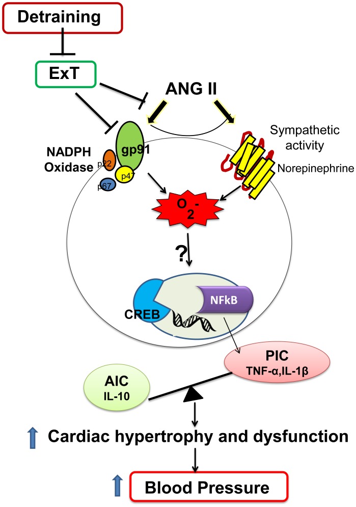
Aims: This study sought to investigate the effects of physical detraining on blood pressure (BP) and cardiac morphology and function in hypertension, and on pro- and anti-inflammatory cytokines (PICs and AIC) and oxidative stress within the brain of hypertensive rats.
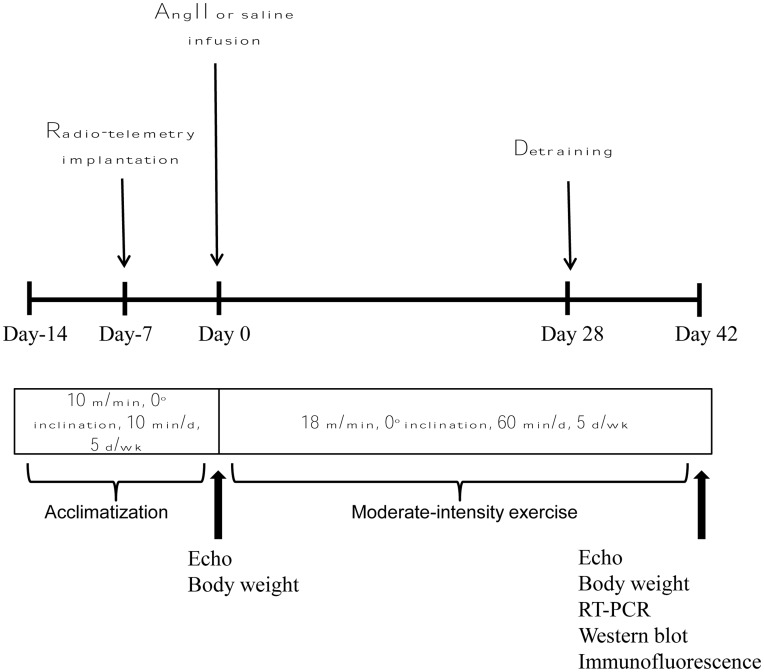
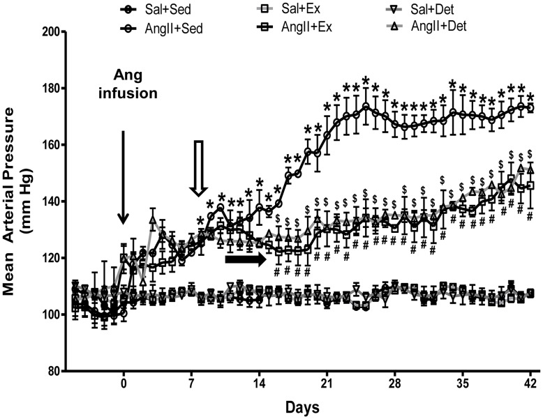
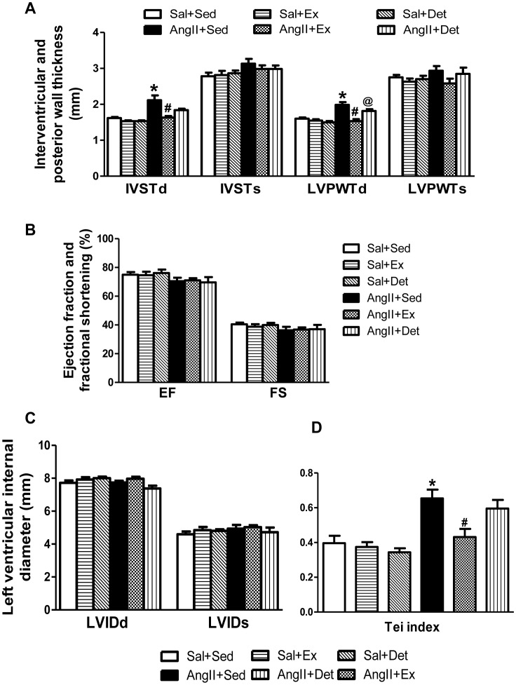
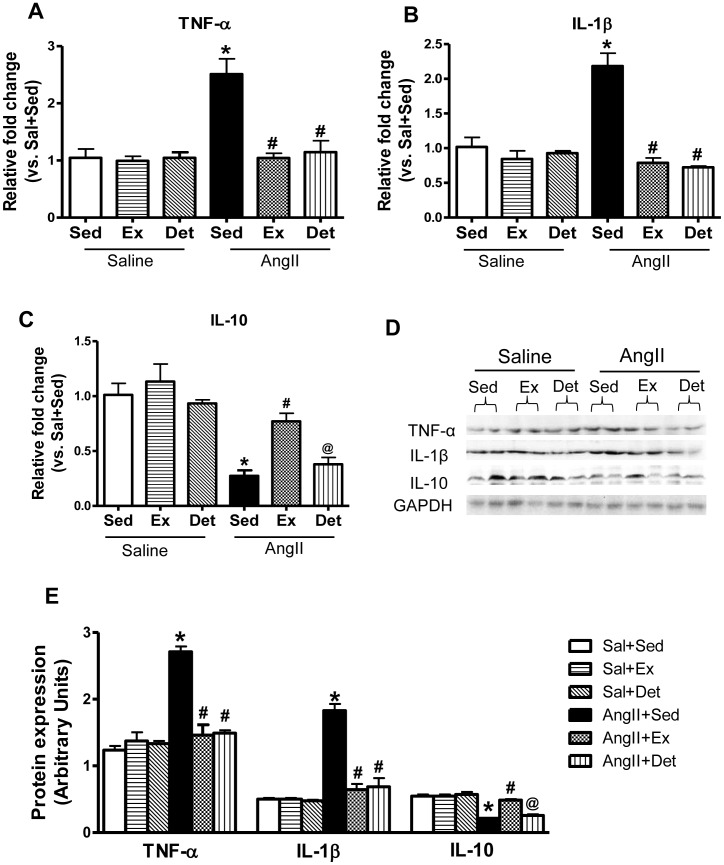
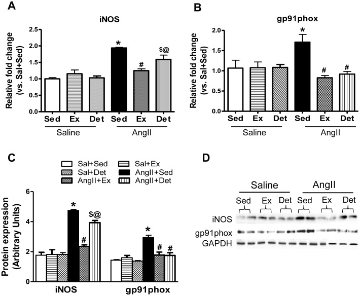
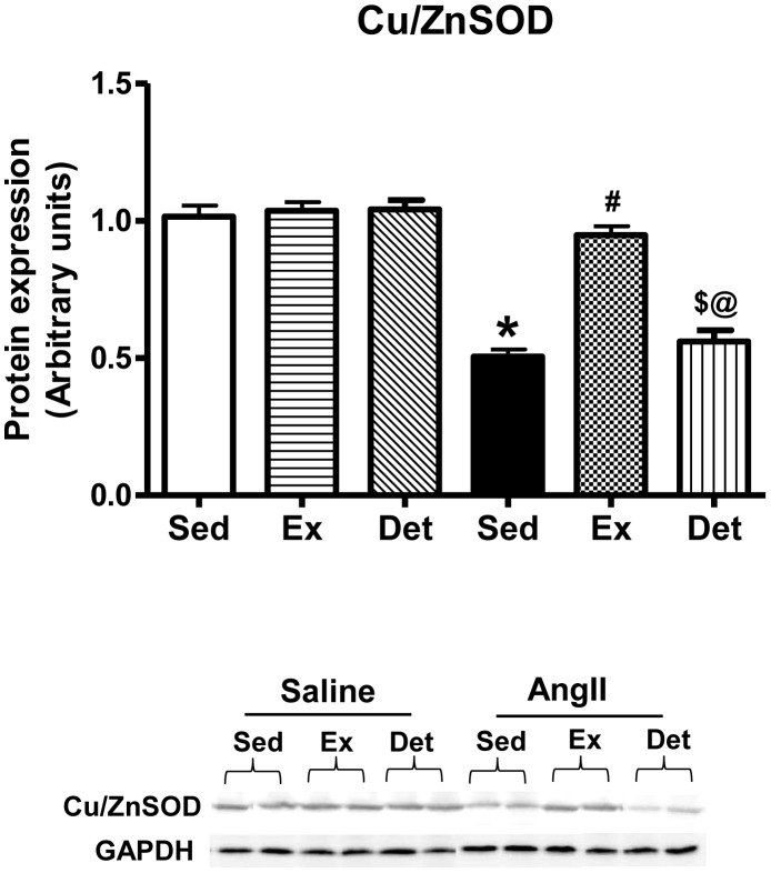
Methods and results: Hypertension was induced in male Sprague-Dawley rats by delivering AngiotensinII for 42 days using implanted osmotic minipumps. Rats were randomized into sedentary, trained, and detrained groups. Trained rats underwent moderate-intensity exercise (ExT) for 42 days, whereas, detrained groups underwent 28 days of exercise followed by 14 days of detraining. BP and cardiac function were evaluated by radio-telemetry and echocardiography, respectively. At the end, the paraventricular nucleus (PVN) was analyzed by Real-time RT-PCR and Western blot. ExT in AngII-infused rats caused delayed progression of hypertension, reduced cardiac hypertrophy, and improved diastolic function. These results were associated with significantly reduced PICs, increased AIC (interleukin (IL)-10), and attenuated oxidative stress in the PVN. Detraining did not abolish the exercise-induced attenuation in MAP in hypertensive rats; however, detraining failed to completely preserve exercise-mediated improvement in cardiac hypertrophy and function. Additionally, detraining did not reverse exercise-induced improvement in PICs in the PVN of hypertensive rats; however, the improvements in IL-10 were abolished.
Conclusion: These results indicate that although 2 weeks of detraining is not long enough to completely abolish the beneficial effects of regular exercise, continuing cessation of exercise may lead to detrimental effects.
Conflict of interest statement
Figures
Similar articles
-
Exercise training attenuates hypertension and cardiac hypertrophy by modulating neurotransmitters and cytokines in hypothalamic paraventricular nucleus.PLoS One. 2014 Jan 17;9(1):e85481. doi: 10.1371/journal.pone.0085481. eCollection 2014. PLoS One. 2014. PMID: 24482680 Free PMC article.
-
Apigenin Improves Hypertension and Cardiac Hypertrophy Through Modulating NADPH Oxidase-Dependent ROS Generation and Cytokines in Hypothalamic Paraventricular Nucleus.Cardiovasc Toxicol. 2021 Sep;21(9):721-736. doi: 10.1007/s12012-021-09662-1. Epub 2021 Jun 2. Cardiovasc Toxicol. 2021. PMID: 34076830
-
Inhibition of NF-κB activity in the hypothalamic paraventricular nucleus attenuates hypertension and cardiac hypertrophy by modulating cytokines and attenuating oxidative stress.Toxicol Appl Pharmacol. 2015 May 1;284(3):315-22. doi: 10.1016/j.taap.2015.02.023. Epub 2015 Mar 7. Toxicol Appl Pharmacol. 2015. PMID: 25759242
-
Beneficial Effects of Exercise on Hypertension-Induced Cardiac Hypertrophy in Adolescents and Young Adults.Curr Hypertens Rep. 2024 Nov;26(11):451-462. doi: 10.1007/s11906-024-01313-4. Epub 2024 Jun 18. Curr Hypertens Rep. 2024. PMID: 38888690 Review.
-
Pathogenetic Mechanisms of Hypertension-Brain-Induced Complications: Focus on Molecular Mediators.Int J Mol Sci. 2022 Feb 23;23(5):2445. doi: 10.3390/ijms23052445. Int J Mol Sci. 2022. PMID: 35269587 Free PMC article. Review.
Cited by
-
Beneficial and harmful effects of exercise in hypertensive patients: the role of oxidative stress.Hypertens Res. 2017 Jan;40(1):15-20. doi: 10.1038/hr.2016.90. Epub 2016 Jul 21. Hypertens Res. 2017. PMID: 27439491 Review.
-
Exercise-Induced Regulation of Redox Status in Cardiovascular Diseases: The Role of Exercise Training and Detraining.Antioxidants (Basel). 2019 Dec 23;9(1):13. doi: 10.3390/antiox9010013. Antioxidants (Basel). 2019. PMID: 31877965 Free PMC article. Review.
-
Combined training in addition to cortisol reduction can improve the mental health of girls with precocious puberty and obesity.Front Pediatr. 2023 Nov 10;11:1241744. doi: 10.3389/fped.2023.1241744. eCollection 2023. Front Pediatr. 2023. PMID: 38027308 Free PMC article.
-
The effects of high-intensity interval training/moderate-intensity continuous training on the inhibition of fat accumulation in rats fed a high-fat diet during training and detraining.Lipids Health Dis. 2024 Jul 22;23(1):221. doi: 10.1186/s12944-024-02209-7. Lipids Health Dis. 2024. PMID: 39039573 Free PMC article.
-
Short-term moderate exercise provides long-lasting protective effects against metabolic dysfunction in rats fed a high-fat diet.Eur J Nutr. 2015 Dec;54(8):1353-62. doi: 10.1007/s00394-014-0816-7. Epub 2014 Dec 21. Eur J Nutr. 2015. PMID: 25528242
References
-
- Coote JH (2005) A role for the paraventricular nucleus of the hypothalamus in the autonomic control of heart and kidney. Exp Physiol 90: 169–173. - PubMed
Publication types
MeSH terms
Substances
Grants and funding
LinkOut - more resources
Full Text Sources
Medical
