

Three-dimensional imaging of Prox1-EGFP transgenic mouse gonads reveals divergent modes of lymphangiogenesis in the testis and ovary
- PMID: 23285114
- PMCID: PMC3527586
- DOI: 10.1371/journal.pone.0052620
Three-dimensional imaging of Prox1-EGFP transgenic mouse gonads reveals divergent modes of lymphangiogenesis in the testis and ovary
Abstract
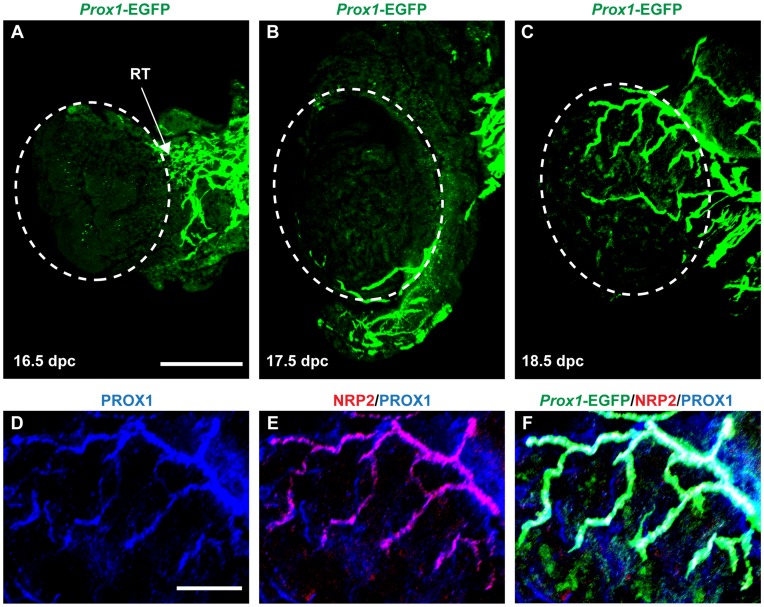
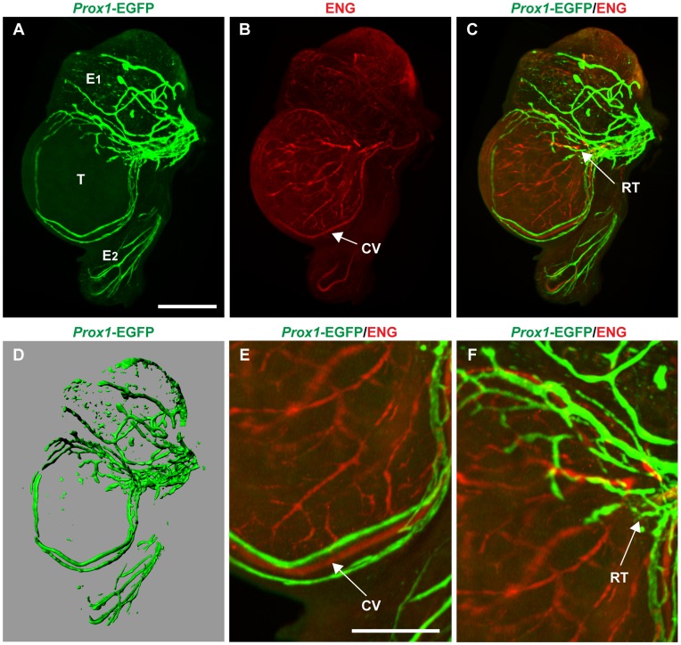
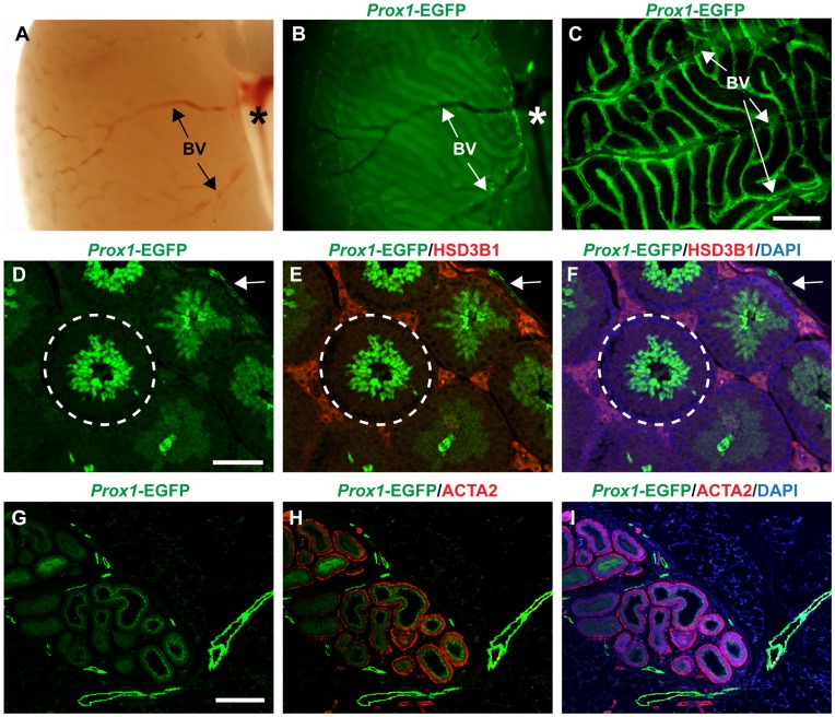
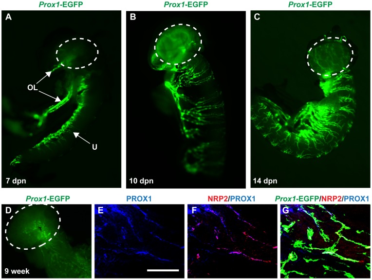
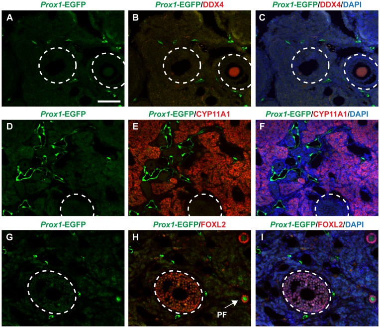
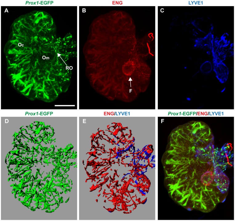
The lymphatic vasculature forms a specialized part of the circulatory system, being essential for maintaining tissue fluid homeostasis and for transport of hormones, macromolecules, and immune cells. Although lymphatic vessels are assumed to play an important role in most tissues, their morphogenesis and function in the gonads remains poorly understood. Here we have exploited a lymphatic-specific Prox1-EGFP reporter mouse model and optical projection tomography technology to characterize both the temporal and spatial development of the lymphatic vessel network in mouse testes and ovaries. We find that lymphangiogenesis in the testis is initiated during late gestation, but in contrast to other organs, lymphatic vessels remain confined to the testis cap and, unlike blood vessels, do not infiltrate the entire organ. Conversely, lymphatic vessels invade the ovarian tissue, beginning postnatally, and sprouting from preexisting lymphatic vessels at the extraovarian rete. The ovary develops a rich network of lymphatic vessels, extending from the medulla into the surrounding cortex adjacent to developing follicles. This study reveals distinct patterns of lymphangiogenesis in the testes and ovaries and will serve as the basis for the identification of the divergent molecular pathways that control morphogenesis and the function of the lymphatic vasculature in these two organs.
Conflict of interest statement
Figures
Similar articles
-
Lymphangiogenesis in mouse embryonic and early postnatal ovaries.Gene Expr Patterns. 2025 Jun;55:119393. doi: 10.1016/j.gep.2025.119393. Epub 2025 Apr 5. Gene Expr Patterns. 2025. PMID: 40194753
-
Visualization of lymphatic vessels by Prox1-promoter directed GFP reporter in a bacterial artificial chromosome-based transgenic mouse.Blood. 2011 Jan 6;117(1):362-5. doi: 10.1182/blood-2010-07-298562. Epub 2010 Oct 20. Blood. 2011. PMID: 20962325 Free PMC article.
-
YAP and TAZ Negatively Regulate Prox1 During Developmental and Pathologic Lymphangiogenesis.Circ Res. 2019 Jan 18;124(2):225-242. doi: 10.1161/CIRCRESAHA.118.313707. Circ Res. 2019. PMID: 30582452
-
Intravital two-photon microscopy of lymphatic vessel development and function using a transgenic Prox1 promoter-directed mOrange2 reporter mouse.Biochem Soc Trans. 2011 Dec;39(6):1674-81. doi: 10.1042/BST20110722. Biochem Soc Trans. 2011. PMID: 22103506 Review.
-
Getting out and about: the emergence and morphogenesis of the vertebrate lymphatic vasculature.Development. 2013 May;140(9):1857-70. doi: 10.1242/dev.089565. Development. 2013. PMID: 23571211 Review.
Cited by
-
Immune and vascular contributions to organogenesis of the testis and ovary.FEBS J. 2022 May;289(9):2386-2408. doi: 10.1111/febs.15848. Epub 2021 Apr 12. FEBS J. 2022. PMID: 33774913 Free PMC article. Review.
-
Cell context-specific expression of primary cilia in the human testis and ciliary coordination of Hedgehog signalling in mouse Leydig cells.Sci Rep. 2015 May 20;5:10364. doi: 10.1038/srep10364. Sci Rep. 2015. PMID: 25992706 Free PMC article.
-
Testicular sex cord-stromal tumors in mice with constitutive activation of PI3K and loss of Pten.Carcinogenesis. 2025 Jan 20;46(1):bgae077. doi: 10.1093/carcin/bgae077. Carcinogenesis. 2025. PMID: 39672957 Free PMC article.
-
Amplification of R-spondin1 signaling induces granulosa cell fate defects and cancers in mouse adult ovary.Oncogene. 2017 Jan 12;36(2):208-218. doi: 10.1038/onc.2016.191. Epub 2016 Jun 6. Oncogene. 2017. PMID: 27270435 Free PMC article.
-
Impact of Excess Activin A on the Lipids, Metabolites, and Steroids of Adult Mouse Reproductive Organs.Endocrinology. 2025 Mar 24;166(5):bqaf054. doi: 10.1210/endocr/bqaf054. Endocrinology. 2025. PMID: 40209098 Free PMC article.
References
-
- Oliver G (2004) Lymphatic vasculature development. Nat Rev Immunol 4: 35–45. - PubMed
-
- Olszewski WL (2003) The lymphatic system in body homeostasis: physiological conditions. Lymphat Res Biol 1: 11–21. - PubMed
-
- Francois M, Caprini A, Hosking B, Orsenigo F, Wilhelm D, et al. (2008) Sox18 induces development of the lymphatic vasculature in mice. Nature 456: 643–647. - PubMed
Publication types
MeSH terms
Substances
LinkOut - more resources
Full Text Sources
Molecular Biology Databases
Miscellaneous
