. 2012;7(12):e52640.
doi: 10.1371/journal.pone.0052640. Epub 2012 Dec 21.

Identification and characterization of FAM124B as a novel component of a CHD7 and CHD8 containing complex

Affiliations

Identification and characterization of FAM124B as a novel component of a CHD7 and CHD8 containing complex

Tserendulam Batsukh et al. PLoS One. 2012.

Abstract

Background: Mutations in the chromodomain helicase DNA binding protein 7 gene (CHD7) lead to CHARGE syndrome, an autosomal dominant multiple malformation disorder. Proteins involved in chromatin remodeling typically act in multiprotein complexes. We previously demonstrated that a part of human CHD7 interacts with a part of human CHD8, another chromodomain helicase DNA binding protein presumably being involved in the pathogenesis of neurodevelopmental (NDD) and autism spectrum disorders (ASD). Because identification of novel CHD7 and CHD8 interacting partners will provide further insights into the pathogenesis of CHARGE syndrome and ASD/NDD, we searched for additional associated polypeptides using the method of stable isotope labeling by amino acids in cell culture (SILAC) in combination with mass spectrometry.

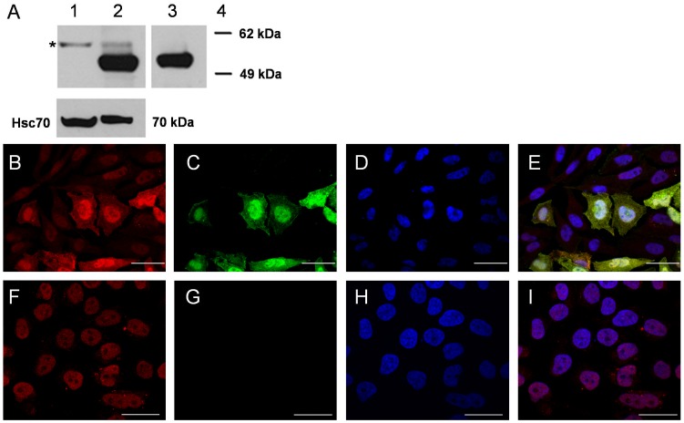
Principle findings: The hitherto uncharacterized FAM124B (Family with sequence similarity 124B) was identified as a potential interaction partner of both CHD7 and CHD8. We confirmed the result by co-immunoprecipitation studies and showed a direct binding to the CHD8 part by direct yeast two hybrid experiments. Furthermore, we characterized FAM124B as a mainly nuclear localized protein with a widespread expression in embryonic and adult mouse tissues.

Conclusion: Our results demonstrate that FAM124B is a potential interacting partner of a CHD7 and CHD8 containing complex. From the overlapping expression pattern between Chd7 and Fam124B at murine embryonic day E12.5 and the high expression of Fam124B in the developing mouse brain, we conclude that Fam124B is a novel protein possibly involved in the pathogenesis of CHARGE syndrome and neurodevelopmental disorders.


Conflict of interest statement

Competing Interests: The authors have declared that no competing interests exist.

Figures

Figure 1
Figure 1. Schematic overview of the SILAC approach.
“Heavy”-labeled cells were co-transfected with the plasmids CHD7-CR1-3-pCMV-HA (containing amino acids 1593-2178, NP_060250.2, in fusion with an HA-tag) and CHD8-pCMV-cmyc (spanning amino acids 1789–2302, NP_065971.2, in fusion with a cmyc-tag). The CHD7 part was purified by anti-HA immunoprecipitation. As a negative control, the same immunoprecipitation was performed in lysates of non-transfected “Light”-labeled HeLa cells. Purified proteins from both cell cultures were pooled in equimolar amounts and in-gel digested, followed by liquid-chromatography (LC)-coupled tandem mass spectrometry.
Figure 2
Figure 2. Schematic overview of FAM124B, CHD7 and CHD8 and the constructs used.
In humans, there are two transcript variants of FAM124B. Transcript variant 1 codes for a protein with 455 amino acids (NP_001116251.1), while transcript variant 2 leads to a shorter protein product with 272 amino acids (NP_079061.2). Both variants have the first 244 amino acids in common. All used FAM124B variant 1 and 2 constructs are full length constructs. Bioinformatic analysis of the amino acid sequence of FAM124B failed to identify any known functional domain, while CHD7 and CHD8 consist of two N-terminal chromodomains (Chromo), followed by a SWI2/SNF2-like ATPase/helicase domain (Helicase/ATPase), three conserved regions (CR1-3), a SANT domain and two BRK domains. The black lines mark the regions cloned into the Yeast two hybrid (pGBKT7, pGADT7) and Co-IP (pCMV-HA, pCMV-cmyc) vectors.
Figure 3
Figure 3. Co-immunoprecipitation of FAM124B with a part of CHD7 and CHD8.
HeLa cells were co-transfected with either the CHD7-CR1-3-pCMV-HA (amino acids 1593-2178, NP_060250.2) plasmid and FAM124B-1,3-pCMV-cmyc or FAM124B-1,3-pCMV-HA (transcript variant 1, NP_001116251.1) or with CHD8-pCMV-cmyc (amino acids 1789-2302, NP_065971.2) and FAM124B-1,3-pCMV-HA (transcript variant 1, NP_001116251.1). (A) Using the anti-CHD8 (abcam, ab84527) or the anti-CHD7 (abcam, ab31824) antibody for precipitation, we detected with the anti-HA antibody (Roche) an approximately 51 kDa band corresponding to the estimated size of FAM124B transcript variant 1. Lane 1: co-transfected Co-IP, lane 2: untransfected HeLa cells as negative control. (B) Reciprocal immunoprecipitation with anti-cmyc antibody (precipitating FAM124B transcript variant 1), and detection with the anti-CHD7 antibody lead to a specific band ∼70 kDa, the estimated size for the CHD7 part fused to the HA-tag. Lane 1: co-transfected Co-IP, lane 2: untransfected HeLa cells as negative control. (C) Reciprocal experiment with anti-HA antibody (precipitating FAM124B transcript variant 1) and detection with the anti-CHD8 antibody detected a specific band ∼68 kDa, the estimated size for the CHD8 part fused to the cmyc-tag. Lane 1: co-transfected Co-IP, lane 2: untransfected HeLa cells as negative control. (D, E, F) The same experimental procedure was performed for FAM124B transcript variant 2, demonstrating a specific interaction of FAM124B transcript variant 2 with the CHD7 and CHD8 part as well. Lane 1: co-transfected Co-IP, lane 2: untransfected HeLa cells as negative control.
Figure 4
Figure 4. Yeast two hybrid assay.
(A) Direct yeast two hybrid experiment with the constructs FAM124B-1,3-pGADT7 (full length transcript variant 1) and CHD7-CR1-3-pGBKT7 (amino acids 1591-2181, NP_060250.2) demonstrating no direct interaction between FAM124B transcript variant 1 and the CHD7 part, while (B) Direct yeast two hybrid experiment with the constructs FAM124B-1,3-pGADT7 (full length transcript variant 1) and CHD8-pGBKT7 (amino acids 1789–2302, NP_065971.2) shows a direct interaction. The same experiments were performed for FAM124B transcript variant 2. (C) Direct yeast two hybrid experiment with the constructs FAM124B-1,2-pGADT7 (full length transcript variant 2) and CHD7-CR1-3-pGBKT7 (amino acids 1591-2181, NP_060250.2). (D) Direct yeast two hybrid experiment with the constructs FAM124B-1,2-pGADT7 (full length transcript variant 2) and CHD8-pGBKT7 (amino acids 1789–2302, NP_065971.2). FAM124B transcript variant 2 interacts directly with the CHD8 part, while no direct interaction with the CHD7 part could be observed.
Figure 5
Figure 5. Western blot and immunocytochemistry of endogenous and overexpressed FAM124B.
(A) Western blot analysis on protein isolated from untransfected HeLa cells (endogenous FAM124B) and HeLa cells overexpressing the FAM124B-1,3-cmyc-tag fusion protein. Lane 1: Immunoblotting of the nuclear cell fraction of untransfected HeLa cells using the FAM124B antibody. The predicted size of human endogenous FAM124B-1,3 is approximately 51 kDa. However, we observed a band of approximately 57 kDa (labeled by an asterisk). We hypothesize that this band could be endogenous FAM124B and the larger size is possibly due to posttranslational modifications of the endogenous protein. Lane 2: Immunoblotting of FAM124B-1,3-pCMV-cmyc overexpressed HeLa total cell lysate using the rabbit anti-FAM124B antibody revealed a prominent band of 51 kDa, the calculated size of human full length FAM124B variant 1 in fusion with a c-myc-tag and the band observed in untransfected HeLa cells. Lane 3: Immunoblotting of the same HeLa cell lysate as shown in lane 2 using anti-c-Myc shows the 51 kDa band corresponding to the overexpressed FAM124B. Lane 4: Marker. Protein quality was controlled using mouse anti-HSC70 producing a 70 kDa band. (B) Immunofluorescence staining using the rabbit anti-Fam124B antibody on HeLa cells transiently transfected with the plasmid FAM124B-1,3-pCMV-cmyc (FAM124B transcript variant 1 fused to an c-myc-tag). Due to the transient transfection not all cells overexpressed FAM124B. Transfected cells show a bright red signal in the nucleus demonstrating a nuclear distribution. The weaker signal in the cell nuclei of the untransfected surrounding cells could possibly reflect an endogenous FAM124B expression, which we detected by RT-PCR and mass spectrometry. (C) Immunofluorescence staining using the anti-c-Myc antibody on the same HeLa cells transiently transfected with the plasmid FAM124B-1,3-pCMV-cmyc showing a bright green signal in the nucleus of the transfected cells. No signal could be observed in the surrounding untransfected cells. (D) Cell nuclei were stained with DAPI (blue). (E) Overlay of B,C and D. (F) Immunofluorescence staining using the rabbit anti-Fam124B antibody on untransfected HeLa cells revealed a weak nuclear signal as it was observed in the surrounding untransfected cells (see Figure 5B). (G) Immunofluorescence staining using the anti-c-Myc antibody on untransfected cells reveals no signal, as expected. (H) Staining of cell nuclei with DAPI (I) overlay of F,G and H. B,C,D,E: Scale bar = 40 µm. F,G,H,I: Scale bar = 30 µm.
Figure 6
Figure 6. Expression pattern of murine Fam124B.
(A) Expression pattern of murine Fam124B by semiquantitative reverse transcription polymerase chain reaction (RT-PCR) on wild type CD1 mouse tissues and E9.5 and E12.5 embryos demonstrating expression in various tissues and during development. (B) Western blot analysis of endogenous Fam124B on various mouse tissues using the anti-FAM124B antibody demonstrates expression in heart, testis, kidney and highest in lung. A weak signal was detected in liver. (C) Immunohistochemistry (IHC) performed on adult mouse tissues slightly counterstained with haematoxylin (blue) confirmed the semiquantitative RT-PCR and western blot results with high expression (brown) in lung, heart, kidney, moderate expression in brain and testis, and very low expression in liver (most liver cell nuclei are only stained with haematoxylin (blue)). Scale bar = 20 µm. Negative = negative control was performed without using the primary antibody.
Figure 7
Figure 7. Fam124B expression in the mouse central nervous system.
(A) Overview, (B) Thalamic nuclei, (C) Hippocampus, (D) Caudate Putamen, (E) Cerebellum. CC = Corpus Callosum, Hipp = Hippocampus, CA1-3 = Cornu Ammonis areas, DG = dentate gyrus, CP = Caudate Putamen, adt = anterior dorsal thalamic nucleus, dg = granual layer of dentate gyrus, lv = lateral ventricle, F = fornix, mdt = mediodorsal thalamic nucleus, IIIv = third ventricle with choroid plexus, vpl = ventral posterior thalamic nucleus, lateral part, ML = molecular layer of cerebellum, PL = purkinje cell layer of cerebellum, GL = granular layer of cerebellum, Scale bar = 100 µm.
Figure 8
Figure 8. In situ hybridization of Fam124B on mouse brain cryosections in comparison with immunostaining.
(A) Immunostaining of a cortex section, (B) In situ hybridization (ISH) of a cortex section (C) Sense control of ISH. (D) Immunostaining of a hippocampus section, (E) In situ hybridization of a hippocampus section (F) Sense control of ISH. DG = dentate gyrus. Scale bar = 100 µm.
Figure 9
Figure 9. Fam124B expression at murine embryonic stage E12.5 (sagittal sections).
Fam124B expression (brown) was found in a variety of embryonic tissues. Expression was observed in several brain areas, spinal cord, dorsal root ganglia, developing cochlea and surrounding tissues, lung, heart, and kidney. Low expression was found in the developing liver and no expression (blue) in blood cells. (A) Overview of Fam124B immunostaining on sagittal section of an E12.5 wildtype embryo slightly counterstained with haematoxylin, scale bar = 200 µm. Higher magnification of (B) Dorsal root ganglia, scale bar = 100 µm (C) Nasal region, scale bar = 100 µm (D) The developing heart, scale bar = 100 µm (E) The developing liver (L), scale bar = 50 µm (F) spinal cord (sc), scale bar = 50 µm (G) The developing lung with segmental bronchus (sb), scale bar = 100 µm (H) Cochlea, and surrounding tissue, scale bar = 100 µm. LV = lateral ventricle, ch.p = choroid plexus differentiating from fourth ventricle, co = cochlea, cd = cochlear duct, ne = nasal epithelium, V gan = left trigeminal (V) ganglion, drg = dorsal root ganglion, VIII gan = Vestibulocochlear (VIII) ganglion, at = left atrium of heart, vt = left heart ventricle, ec = endothelial cells, car = cartilage primordium of body of vertebra, ccp = cartilage condensation being primordium of vertebral body.

Similar articles

Cited by

References

    1. Vissers LE, van Ravenswaaij CM, Admiraal R, Hurst JA, de Vries BB, et al. (2004) Mutations in a new member of the chromodomain gene family cause CHARGE syndrome. Nat Genet 36: 955–957. - PubMed
    1. Aramaki M, Udaka T, Kosaki R, Makita Y, Okamoto N, et al. (2006) Phenotypic spectrum of CHARGE syndrome with CHD7 mutations. J Pediatr 148: 410–414. - PubMed
    1. Jongmans MC, Admiraal RJ, van der Donk KP, Vissers LE, Baas AF, et al. (2006) CHARGE syndrome: the phenotypic spectrum of mutations in the CHD7 gene. J Med Genet 43: 306–314. - PMC - PubMed
    1. Lalani SR, Safiullah AM, Fernbach SD, Harutyunyan KG, Thaller C, et al. (2006) Spectrum of CHD7 mutations in 110 individuals with CHARGE syndrome and genotype-phenotype correlation. Am J Hum Genet 78: 303–314. - PMC - PubMed
    1. Sanlaville D, Etchevers HC, Gonzales M, Martinovic J, Clement-Ziza M, et al. (2006) Phenotypic spectrum of CHARGE syndrome in fetuses with CHD7 truncating mutations correlates with expression during human development. J Med Genet 43: 211–217. - PMC - PubMed

Publication types

MeSH terms