. 2012;7(12):e52648.
doi: 10.1371/journal.pone.0052648. Epub 2012 Dec 20.

Genotyping of fanconi anemia patients by whole exome sequencing: advantages and challenges

Affiliations

Genotyping of fanconi anemia patients by whole exome sequencing: advantages and challenges

Kerstin Knies et al. PLoS One. 2012.

Abstract

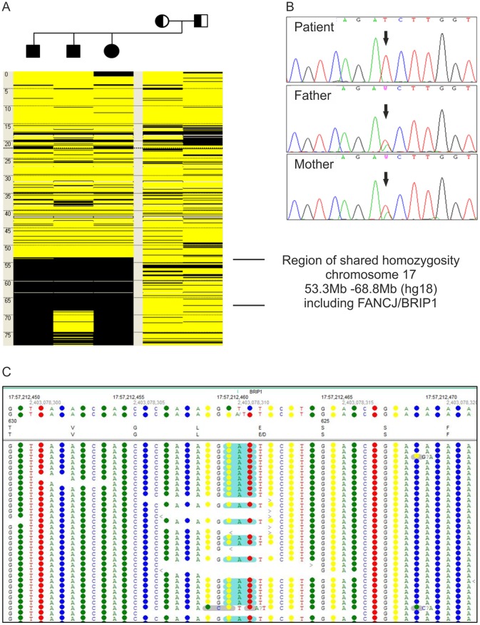
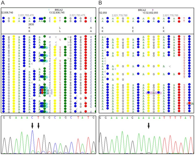
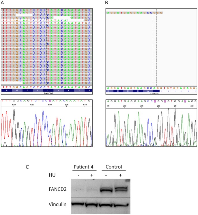
Fanconi anemia (FA) is a rare genomic instability syndrome. Disease-causing are biallelic mutations in any one of at least 15 genes encoding members of the FA/BRCA pathway of DNA-interstrand crosslink repair. Patients are diagnosed based upon phenotypical manifestations and the diagnosis of FA is confirmed by the hypersensitivity of cells to DNA interstrand crosslinking agents. Customary molecular diagnostics has become increasingly cumbersome, time-consuming and expensive the more FA genes have been identified. We performed Whole Exome Sequencing (WES) in four FA patients in order to investigate the potential of this method for FA genotyping. In search of an optimal WES methodology we explored different enrichment and sequencing techniques. In each case we were able to identify the pathogenic mutations so that WES provided both, complementation group assignment and mutation detection in a single approach. The mutations included homozygous and heterozygous single base pair substitutions and a two-base-pair duplication in FANCJ, -D1, or -D2. Different WES strategies had no critical influence on the individual outcome. However, database errors and in particular pseudogenes impose obstacles that may prevent correct data perception and interpretation, and thus cause pitfalls. With these difficulties in mind, our results show that WES is a valuable tool for the molecular diagnosis of FA and a sufficiently safe technique, capable of engaging increasingly in competition with classical genetic approaches.


Conflict of interest statement

Competing Interests: The authors have declared that no competing interests exist.

Figures

Figure 1
Figure 1. Cell cycle analysis.
(A) Graphic presentation of the results of flow cytometric cell cycle analysis. Peripheral blood lymphocytes were exposed to MMC. The ratio “sum of all G2 phases vs. growth fraction” was calculated from individual cultures and plotted against the G0/G1 phase compartment. Cells from patient 1 and the siblings 2–1, 2–2 and 2–3 show high ∑G2/GF ratios (blue squares) similar to those from other persons with FA (red diamonds), but were distinct of normal controls (green dots). (B) Flow histograms of fibroblast cultures from patients 3 and 4 show increased G2 arrest after exposure to MMC, in contrast to a control cell line (arrows).
Figure 2
Figure 2. Genotyping of patient 1.
(A) The heterozygous FANCD2 splice site mutation c.3888+2T>G in patient 1. The upper panel demonstrates mutation calling in NGS data format. The lower panel shows an electropherogram of Sanger cDNA sequencing depicting heterozygous skipping of exon 39. (B) The heterozygous FANCD2 missense mutation c.2204G>A. The upper panel demonstrates the substitution in NGS data format, while the lower panel shows the confirmation by Sanger sequencing of gDNA. (C) An immunoblot shows very faint FANCD2-S and -L bands after exposure of fibroblasts from patients 1 to MMC (lane 3). This was similar to other FA-D2 patients (example on lane 2) but contrasted markedly to normal controls (example on lane 1). RAD50 was used as loading control.
Figure 3
Figure 3. Genotyping of patient 2.
(A) Homozygous mutation call c.1878A>T in FANCJ detected in NGS data of patient 2–1. (B) Autozygosity mapping with SNP data of the family of project 2. The figure schematically presents chromosome 17 (positions in Mb) of each family member. Heterozygous SNP calls are displayed in yellow, homozygous calls in black. The three affected siblings share a homozygous region between 53.3 Mb and 68.8 Mb. (C) Confirmation of homozygosity of the mutation in patient 2–1 and heterozygosity in his parents by Sanger sequencing electropherograms, consistent with Mendelian segregation.
Figure 4
Figure 4. Genotyping of patient 3.
(A) NGS data in the upper panel show the insertion c.7890_7891insAA in FANCD1 detected in patient 3. The electropherogram in the lower panel demonstrates corresponding validation by Sanger sequencing. (B) The upper panel misleadingly displays the single-bp substitution c.7795G>A in the NGS data of patient 3 as a SNP, highlighted in pink. Confirmation by Sanger sequencing is shown below.
Figure 5
Figure 5. Genotyping of patient 4.
(A) Displayed is the FANCD2 mutation c.1370T>C in patient 4 in NGS data as well as validated by Sanger sequencing. (B) The upper panel shows NGS data with low coverage of FANCD2 exon 5 containing the substitution c.376A>G. The electropherogram in the lower panel depicts validation by Sanger sequencing. (C) Hydroxyurea (HU) treated (+) and untreated (−) fibroblasts of patient 4 show very low levels of both the S and L species of residual FANCD2 protein. Vinculin was used as loading control.

References

    1. Vuorela M, Pylkas K, Hartikainen JM, Sundfeldt K, Lindblom A, et al. (2011) Further evidence for the contribution of the RAD51C gene in hereditary breast and ovarian cancer susceptibility. Breast Cancer Res Treat 130: 1003–1010. - PubMed
    1. Alter BP, Kupfer G (1993) Fanconi Anemia. In: Pagon RA, Bird TD, Dolan CR, Stephens K, editors. GeneReviews. Seattle (WA). - PubMed
    1. Auerbach AD (2009) Fanconi anemia and its diagnosis. Mutat Res 668: 4–10. - PMC - PubMed
    1. Seif AE (2011) Pediatric leukemia predisposition syndromes: clues to understanding leukemogenesis. Cancer Genet 204: 227–244. - PubMed
    1. Rosenberg PS, Greene MH, Alter BP (2003) Cancer incidence in persons with Fanconi anemia. Blood 101: 822–826. - PubMed

Publication types

Substances