. 2012;7(12):e52684.
doi: 10.1371/journal.pone.0052684. Epub 2012 Dec 28.

Diverged alleles of the Anopheles gambiae leucine-rich repeat gene APL1A display distinct protective profiles against Plasmodium falciparum

Affiliations

Diverged alleles of the Anopheles gambiae leucine-rich repeat gene APL1A display distinct protective profiles against Plasmodium falciparum

Inge Holm et al. PLoS One. 2012.

Abstract

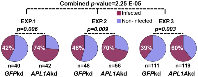
Functional studies have demonstrated a role for the Anopheles gambiae APL1A gene in resistance against the human malaria parasite, Plasmodium falciparum. Here, we exhaustively characterize the structure of the APL1 locus and show that three structurally different APL1A alleles segregate in the Ngousso colony. Genetic association combined with RNAi-mediated gene silencing revealed that APL1A alleles display distinct protective profiles against P. falciparum. One APL1A allele is sufficient to explain the protective phenotype of APL1A observed in silencing experiments. Epitope-tagged APL1A isoforms expressed in an in vitro hemocyte-like cell system showed that under assay conditions, the most protective APL1A isoform (APL1A(2)) localizes within large cytoplasmic vesicles, is not constitutively secreted, and forms only one protein complex, while a less protective isoform (APL1A(1)) is constitutively secreted in at least two protein complexes. The tested alleles are identical to natural variants in the wild A. gambiae population, suggesting that APL1A genetic variation could be a factor underlying natural heterogeneity of vector susceptibility to P. falciparum.


Conflict of interest statement

Competing Interests: The authors have declared that no competing interests exist.

Figures

Figure 1
Figure 1. Genomic organisation of the A. gambiae APL1 region.
The genomic region of the 2L chromosome that comprises the APL1 locus is indicated by a green bar (A. gambiae reference genome , genome assembly AgamP3). The bar delimits the region of the genome that has been completely spanned with overlapping PCR amplicons. The chromosomal positions are indicated (in bp) on top of the bar. The yellow bars indicate the regions for which overlapping PCR fragments have been sequenced. Blue arrows represent the genes in this chromosomal region and their direction of transcription. The last two lines indicate gene identifiers and names.
Figure 2
Figure 2. Alignment and comparison of the Ngousso APL1A alleles. A)
Schematic view of the genomic regions of the different APL1A alleles identified in A. gambiae Ngousso. Each row represents the genomic region of one APL1A allele. The first row shows the APL1A gene from the A. gambiae reference genome. Each of the following three rows represents one of the APL1A alleles present in the Ngousso strain, APL1A1, APL1A2, APL1A3, respectively. The sequences belonging to the APL1A genes are represented as solid bars with white boxes corresponding to the 5′ non-coding region and the region of the first intron. Dashed lines illustrate introduced gaps for a better alignment of the different alleles. DNA regions adjacent of the APL1A genes are shown as black horizontal lines. Predicted peptide domains are indicated as follows: signal peptide (black vertical bar), additional sequences and PANGGL repeat region (yellow), LRR repeat region (green), coiled-coil domain (orange). Yellow triangles indicate the positions and length of indels, red stars indicate the positions of the translation stop codons. At the bottom right, the black bar indicates the DNA region targeted by the dsRNA. B) Schematic representation of the labelled PstI-RFLP fragments of APL1A alleles obtained by the high-throughput RFLP genotyping assay in Ngousso. The first line represents the genomic DNA of this region with two arrows marking the positions of the forward and reverse fluorescent-labeled primers used in the assay. The red and the blue vertical bars indicate the respective position of the terminal PstI restriction sites in the different alleles. The fragment sizes of the labelled PstI fragment generated from the 5′ end of the Ngousso APL1A alleles are indicated in red. The sizes of the labelled PstI fragment from the 3′ end of the APL1A alleles are in blue.
Figure 3
Figure 3. Schematic representation of the three different APL1 haplotypes found in A. gambiae Ngousso.
Each row shows one of the three identified APL1 haplotypes of Ngousso, numbered 1 to 3. The three APL1 proteins encoded by each haplotype are shown. Predicted peptide domains are indicated as follows: black vertical bar – signal sequence, yellow box – PANGGL repeat region, green – LRR repeat region, orange – coiled-coil domain.
Figure 4
Figure 4. APL1A silencing renders mosquitoes significantly more susceptible to P. falciparum infection.
Silencing of APL1A in A. gambiae Ngousso results in significantly higher infection prevalence in mosquitoes fed on in vitro cultured P. falciparum gametocytes. For each experiment (EXP.), a Chi-square analysis was performed to compare infection prevalence between GFP-knockdown (GFPkd) and APL1Akd. A meta-analysis using the Fisher method was also used to combine the p-values of the three independent experiments.
Figure 5
Figure 5. Differential secretion pattern and sub-cellular localization of APL1A-V5 alleles in hemocyte-like cell culture. A)
Immunofluorescence analysis of 4a-3A hemocyte-like cells transfected with plasmids encoding V5-tagged APL1A1, APL1A2 and APL1A3. Cells were stained with Hoechst 33342 to label nuclei (blue). Staining with a mouse anti-V5 mAb followed by anti-mouse Alexa 488-conjugated IgG (red) indicates that APL1A1 exhibits a diffuse pattern in the cytoplasm whereas APL1A2 and APL1A3 are essentially localized in vesicles. Pictures were taken under equivalent exposure conditions. B) Immunoblot analysis of cells (C) and culture medium (M) of the 4a-3A hemocyte-like cell line transfected with plasmids encoding V5-tagged APL1A1, APL1A2 and APL1A3 under reducing (R) and non-reducing (NR) conditions. Immunoblots were probed with a mouse anti-V5 mAb, protein quantities on the different blots are not comparable. APL1A1 is secreted in the culture medium as at least two protein complexes under non-reducing conditions, whereas APL1A2 and APL1A3 are retained in the cell cytoplasm and form only one protein complex under non-reducing conditions. Estimated sizes of monomeric APL1A forms including V5-tag are: 76 kDa (APL1A1), 60 kDa (APL1A2) and 51 kDa (APL1A3). C) Amino acid alignment of cysteine-rich regions of the three Ngousso APL1A proteins and A. gambiae PEST APL1C. Numbers correspond to the amino acid positions. Blue stars represent stop codons, cysteine residues are highlighted in red. The cysteine residue corresponding to the position 562 in the APL1C sequence (APL1CC562, black arrow) described by Povelones et al. , is involved in the disulfide-linked complex formed between LRIM1 and APL1C and is only conserved in APL1A1. The same cysteine is also referred to as APL1C C551 in the APL1C protein published by Baxter et al. .

References

    1. TDR/WHO (2012) Malaria.
    1. Niare O, Markianos K, Volz J, Oduol F, Toure A, et al. (2002) Genetic loci affecting resistance to human malaria parasites in a West African mosquito vector population. Science 298: 213–216. - PubMed
    1. Menge DM, Zhong D, Guda T, Gouagna L, Githure J, et al. (2006) Quantitative trait loci controlling refractoriness to Plasmodium falciparum in natural Anopheles gambiae mosquitoes from a malaria-endemic region in western Kenya. Genetics 173: 235–241. - PMC - PubMed
    1. Riehle MM, Markianos K, Niare O, Xu J, Li J, et al. (2006) Natural malaria infection in Anopheles gambiae is regulated by a single genomic control region. Science 312: 577–579. - PubMed
    1. Riehle MM, Markianos K, Lambrechts L, Xia A, Sharakhov I, et al. (2007) A major genetic locus controlling natural Plasmodium falciparum infection is shared by East and West African Anopheles gambiae. Malar J 6: 87. - PMC - PubMed

Publication types