. 2012;7(12):e52700.
doi: 10.1371/journal.pone.0052700. Epub 2012 Dec 28.

Functional comparison of chronological and in vitro aging: differential role of the cytoskeleton and mitochondria in mesenchymal stromal cells

Affiliations

Functional comparison of chronological and in vitro aging: differential role of the cytoskeleton and mitochondria in mesenchymal stromal cells

Sven Geissler et al. PLoS One. 2012.

Abstract

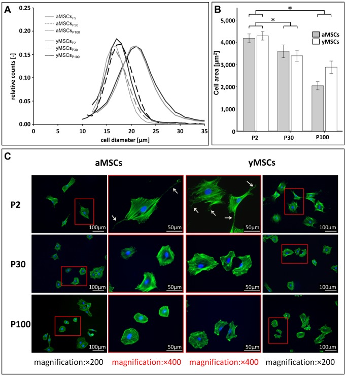
Mesenchymal stromal cells (MSCs) are of high relevance for the regeneration of mesenchymal tissues such as bone and cartilage. The promising role of MSCs in cell-based therapies and tissue engineering appears to be limited due to a decline of their regenerative potential with increasing donor age, their limited availability in human tissues and the need of in vitro expansion prior to treatment. We therefore aimed to determine to which degree in vitro aging and chronological aging may be similar processes or if in vitro culture-related changes at the cellular and molecular level are at least altered as a function of donor age. For that purpose we established MSCs cultures from young (yMSCs) and aged (aMSCs) rats that were cultured for more than 100 passages. These long-term MSCs cultures were non-tumorigenic and exhibited similar surface marker patterns as primary MSCs of passage 2. During in vitro expansion, but not during chronological aging, MSCs progressively lose their progenitor characteristics, e.g., complete loss of osteogenic differentiation potential, diminished adipogenic differentiation, altered cell morphology and increased susceptibility towards senescence. Transcriptome analysis revealed that long-term in vitro MSCs cultivation leads to down-regulation of genes involved in cell differentiation, focal adhesion organization, cytoskeleton turnover and mitochondria function. Accordingly, functional analysis demonstrated altered mitochondrial morphology, decreased antioxidant capacities and elevated ROS levels in long-term cultivated yMSCs as well as aMSCs. Notably, only the MSC migration potential and their antioxidative capacity were altered by in vitro as well as chronological aging. Based on specific differences observed between the impact of chronological and in vitro MSC aging we conclude that both are distinct processes.


Conflict of interest statement

Competing Interests: The authors have declared that no competing interests exist.

Figures

Figure 1
Figure 1. Generation and characterization of in vitro aged MSCs.
(A): Cumulative population doublings of aMSCs and yMSCs during the first 80 days of culture are shown (n = 5). (B): Long-term cultivation has no influence on short-term proliferation rate of aMSCs and yMSCs of passage 30 and 100. Proliferation assay was performed using CyQuant®. (C): Graphs illustrate quantified signal intensities of p21WAF1/CIP1 and p16INK4A relative to GAPDH. (D): Representative Western blots showing increased p21WAF1/CIP1 and p16INK4A expression during in vitro aging. GAPDH served as endogenous control. (E): In anchorage-independent growth assays in vitro aged MSCsP100 did not form colonies, while the breast carcinoma cell line MDA-MB-231, which served as positive control, produced numerous colonies (n = 3). Abbreviations: aMSCs, mesenchymal stromal cells from aged donors; yMSCs, mesenchymal stromal cells from young donors; P: passage. * indicates statistical significance (p<0.05).
Figure 2
Figure 2. Long-term in vitro culture alters MSC morphology independent from the donor age.
(A): Cell diameter of aMSCs and yMSCs decreases during the course of long-term cultivation. Diagram shows the cell size distribution of MSCs measured by CASY® TT cell analyzer system at indicated passages after trypsinization. (B): Cellular area of attached aMSCs and yMSCs significantly decreases during in vitro aging. Measurements were performed from fluorescence images of identical exposure conditions. (C): Representative images of phalloidin labeled MSCs highlight reduction of cellular expansion. Additionally, in vitro aged aMSCs and yMSCs exhibited less filopodia, lamellipodia and cell spreading (white arrows). * indicates statistical significance (p<0.05).
Figure 3
Figure 3. Long-term cultivation negatively influences the differentiation and migration potential of aMSCs and yMSCs.
(A): In contrast to primary MSCs of passage 2, in vitro aged aMSCs and yMSCs of P30 and P100 show no matrix mineralization. Osteogenic differentiation was initiated with dexamethason and determined by matrix mineralization (Alizarin Red, AR) and normalized to cell number (alamarBlue®, AB). Dashed lines indicate differentiation potential of the negative control cultured in EM. (B): Under stimulation with BMP2, aMSCs and yMSCs of P2 show strong osteogenic differentiation, while again no matrix mineralization was observed in long-term MSC cultures of P30 and P100. (C): Adipogenic differentiation of aMSCs and yMSCs of P30 and P100, induced by adipogenic medium, was diminished by 50% compared to aMSCs and yMSCs of P2. In reference to the negative control maintained in EM (dashed line), aMSCs and yMSCs of P30 and P100 retained a potential for adipogenic differentiation. Differentiation was determined by using Oil red O (OR) staining and normalized to cell number. Diagram shows values normalized to negative control. (D): The number of migrated cells declined with increased in vitro passage. Moreover, aMSCs of each passage demonstrated significantly lower migratory potential compared to yMSCs. Migration rates were measured with a modified Boyden chamber assay. At least five independent experiments were carried out for all assays. Abbreviations: OD, optical density. * indicates statistical significance (p<0.05).
Figure 4
Figure 4. Transcriptional profiling of aMSCs and yMSCs at P2, P30 and P100.
(A): The absolute number of genes detected after thresholding diminished during advanced in vitro culture independent from donor age (second column). The correlation coefficient (r2) was significantly reduced between aMSCs and yMSCs of P30 and P100 compared to P2. Only minor differences in gene expression were detected between aMSCs and yMSCs of each passage. (B): Functional annotation clustering of genes exclusively expressed either in primary MSC of P2 or in vitro aged MSCs of P30 and P100 revealed 431 and 124 differentially regulated genes, respectively. At P2 genes were mainly associated with chemokine signaling, apoptosis, cell migration, and calcium homeostasis. Whereas at P30 and P100 exclusively expressed genes are involved in Notch signaling, cell cycle progression and receptor signaling. (C): Analysis of pathways down-regulated after long-term in vitro culture revealed involvement of mitochondria, focal adhesions, cytoskeleton organization, TGF-β/BMP, WNT, and PPARγ signaling. Pathways up-regulated upon long-term in vitro culture were associated with cell cycle progression, DNA replication, p53, MAPK, and insulin signaling. (D): Differential statistical analysis summarizes all pathways and genes significantly up- and down-regulated during in vitro culture. The most numerous genes down-regulated during in vitro aging of aMSCs and yMSCs were associated with focal adhesions, actin cytoskeleton organization and mitochondrial function.
Figure 5
Figure 5. Long-term cultivation of MSCs alters their mitochondrial function.
(A): Fluorescence microscopy was used to investigate the morphology of the mitochondrial network within long-term cultivated and primary aMSCs and yMSCs. Upon in vitro aging mitochondrial network appeared to be altered. Images show immunofluorescence of mitochondria and the actin cytoskeleton stained with a specific antibody recognizing cytochrome C and Alexa 594-conjugated phalloidin, respectively. Nuclei were counterstained with DAPI. (B): During in vitro aging the relative mitochondrial area per cell area increases in aMSCs and yMSCs of passage P30 and P100 compared to P2. The mitochondrial network and the cellular area were quantified after staining with MitoTracker™ Red and phalloidin, respectively. Diagram values represent ratio of the mitochondria network area relative to the cell area. (C): The total antioxidant capacity decreases with increasing passage number. Moreover, yMSCs of P2 and P100 exhibited significant higher antioxidant activities than aMSCs of the same passage. The Trolox® equivalent antioxidant assay kit was used to determine the total antioxidant capacity of whole MSC lysates and quantified against a Trolox® standard row. (D): Intracellular ATP levels decline significantly in long-term cultivated aMSCs and yMSCs of passage P30 and P100. Cellular ATP was determined using ATPLite™ bioluminescence luciferase-based assay and normalized to total DNA content determined by CyQuant®. (E): Long-term cultivated yMSCs and aMSCs of P30 and P100 displayed higher ROS production than primary MSCs of P2. After treatment with pyocyanin, which increases ROS levels, the observed difference between P2 and P30/P100 remained but the absolute value amplified about 2-fold. Intracellular ROS level were determined using CM-H2-DCFDA and normalized to total DNA content. (F): Measurement of the mitochondrial membrane potential (ΔΨm) revealed a progressive increase during in vitro aging with highest values in aMSCs of P100. Upon treatment with valomycin, an inhibitor of the mitochondrial respiratory chain, ΔΨm declined in aMSCs and yMSCs of all passages. The mitochondrial ΔΨm was determined with the MitoProbe® JC-1. * indicates statistical significance (p<0.05).

References

    1. Salem HK, Thiemermann C (2010) Mesenchymal stromal cells: current understanding and clinical status. Stem Cells 28: 585–596. - PMC - PubMed
    1. Kasper G, Glaeser JD, Geissler S, Ode A, Tuischer J, et al. (2007) Matrix metalloprotease activity is an essential link between mechanical stimulus and mesenchymal stem cell behavior. Stem Cells 25: 1985–1994. - PubMed
    1. Kasper G, Dankert N, Tuischer J, Hoeft M, Gaber T, et al. (2007) Mesenchymal stem cells regulate angiogenesis according to their mechanical environment. Stem Cells 25: 903–910. - PubMed
    1. Krampera M, Pizzolo G, Aprili G, Franchini M (2006) Mesenchymal stem cells for bone, cartilage, tendon and skeletal muscle repair. Bone 39: 678–683. - PubMed
    1. Le Blanc K, Pittenger M (2005) Mesenchymal stem cells: progress toward promise. Cytotherapy 7: 36–45. - PubMed

Publication types