. 2012;7(12):e52754.
doi: 10.1371/journal.pone.0052754. Epub 2012 Dec 28.

Cancer progression mediated by horizontal gene transfer in an in vivo model

Affiliations

Cancer progression mediated by horizontal gene transfer in an in vivo model

Catalina Trejo-Becerril et al. PLoS One. 2012.

Abstract

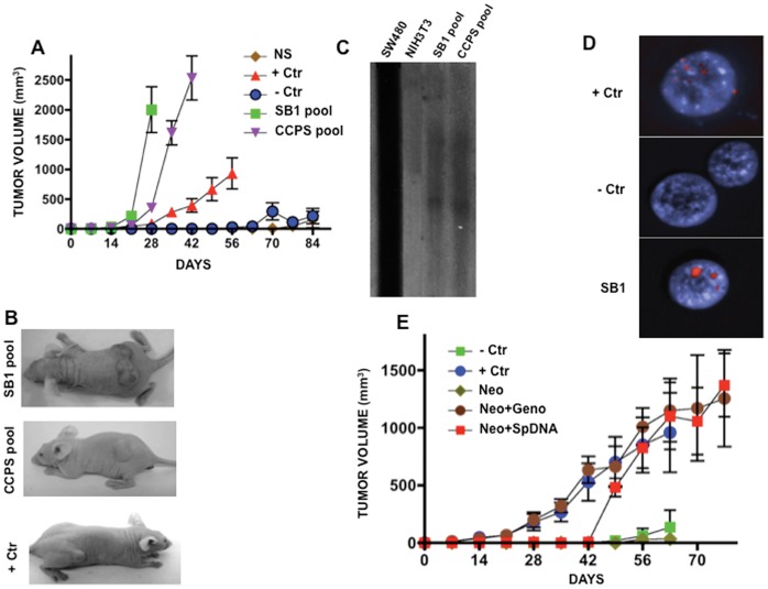
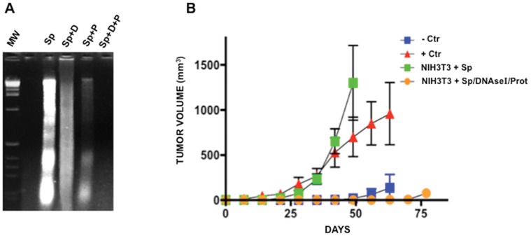
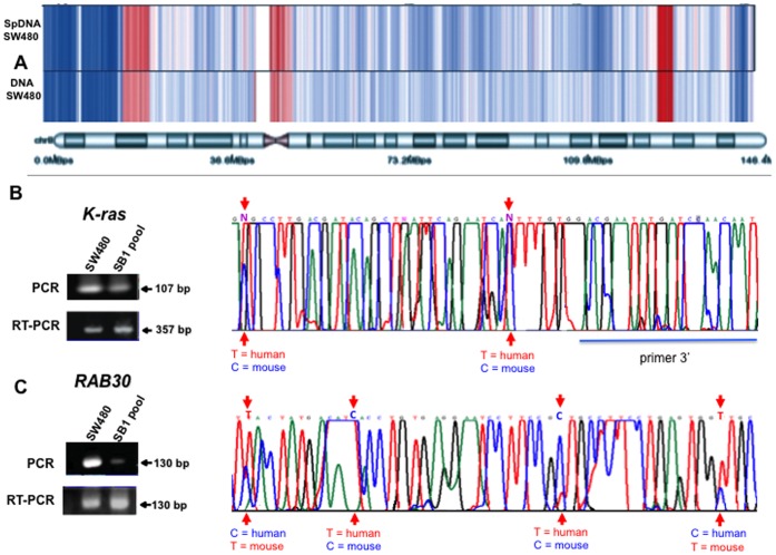
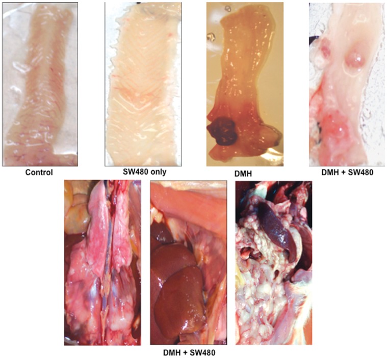
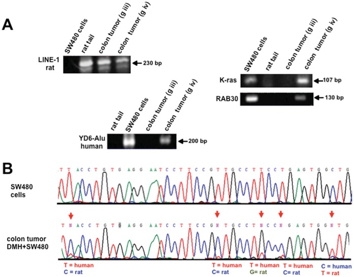
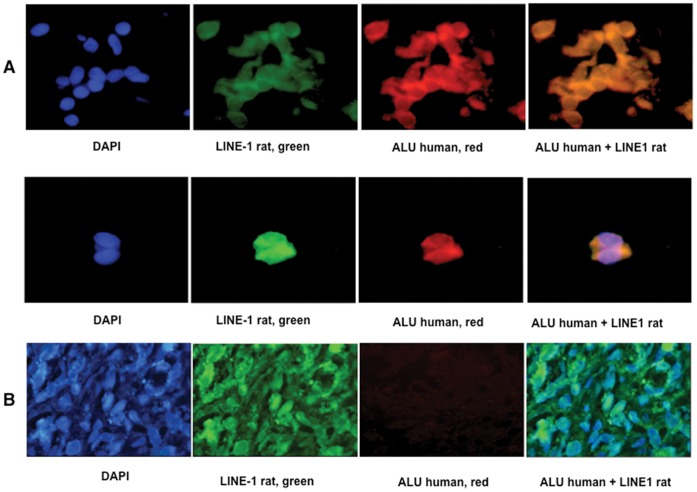
It is known that cancer progresses by vertical gene transfer, but this paradigm ignores that DNA circulates in higher organisms and that it is biologically active upon its uptake by recipient cells. Here we confirm previous observations on the ability of cell-free DNA to induce in vitro cell transformation and tumorigenesis by treating NIH3T3 recipient murine cells with serum of colon cancer patients and supernatant of SW480 human cancer cells. Cell transformation and tumorigenesis of recipient cells did not occur if serum and supernatants were depleted of DNA. It is also demonstrated that horizontal cancer progression mediated by circulating DNA occurs via its uptake by recipient cells in an in vivo model where immunocompetent rats subjected to colon carcinogenesis with 1,2-dimethylhydrazine had increased rate of colonic tumors when injected in the dorsum with human SW480 colon carcinoma cells as a source of circulating oncogenic DNA, which could be offset by treating these animals with DNAse I and proteases. Though the contribution of biologically active molecules other than DNA for this phenomenon to occur cannot be ruled out, our results support the fact that cancer cells emit into the circulation biologically active DNA to foster tumor progression. Further exploration of the horizontal tumor progression phenomenon mediated by circulating DNA is clearly needed to determine whether its manipulation could have a role in cancer therapy.


Conflict of interest statement

Competing Interests: The authors have the following interest. Co-author Phillipe Anker is affiliated to OncoXL. There are no patents, products in development or marketed products to declare. This does not alter the authors’ adherence to all the PLOS ONE policies on sharing data and materials, as detailed online in the guide for authors.

Figures

Figure 1
Figure 1. Tumorigenesis and DNA transfer in recipient murine NIH3T3 cells after passive transfection. A.
Tumor growth in nude mice from “passively” transformed cells. Faster and higher tumor growth was observed in SB1 pool and CCPS pool (NIH3T3 exposed to supernatant of SW480 cells and to the serum of a patient with colon cancer, respectively). SW480 cells were used as positive control, whereas NIH3T3 and NIH3T3 exposed to normal serum showed essentially no growth. B. Representative pictures of tumors in mice from each group. C. Southern blot hybridization of SB1 and CCPS pools of cells against genomic DNA of SW480 cells. Lane SW480 cells are the positive control and NIH3T3, the negative one. A clear hybridization signal is only observed in SB1 and CCPS lanes. D. FISH analysis of repetitive human sequences. Positive control is human lymphocytes and murine cells negative control. SB1 cells shows strong signal. E. Tumor growth is similar in NIH3T3 actively transfected with genomic DNA from SW480 cells (Neo-Geno) and actively transfected DNA extracted from supernatant of SW480 cells as compared with no growth in NIH3T3 (-Crt) and transfected with the empty-vector only. Positive control, SW480 cells.
Figure 2
Figure 2. Tumorigenesis after “active” transfection with DNA supernatant of SW480 cells and passive transfection using DNA-depleted supernatant. A.
Agarose gel electrophoresis of DNA extracted from supernatant (Sp), DNA from supernatant treated with DNAse I (Sp+D), protease only (Sp+P), and both (Sp+D+P). DNA is partially degraded by DNAse I and protease, but fully degraded when exposed to both treatments. B. No tumor growth was observed in NIH3T3 exposed to DNA-depleted (DNAse I/Prot) supernatant of SW480 cells, while passively transfected NIH3T3 with untreated supernatant are tumorigenic. +Ctr are SW480 cells and −Ctr are NIH3T3 cells.
Figure 3
Figure 3. DNA copy number analysis of extracellular and intracellular DNA from SW480 cells and gene transfer to murine NIH3T3 cells. A.
Heat map representing the DNA copy number along chromosome 8. Blue represents regions with deletions and red regions with amplifications. A nearly identical pattern of DNA copy number changes between extracellular (SpDNA SW480) and intracellular (DNA SW480) DNA, compared to a common normal reference can be observed. RT-PCR, PCR and sequencing of Human K-ras (B) and RAB30 (C). Negative control was NIH3T3 cells.
Figure 4
Figure 4. Tumorigenesis in the immunocompetent Wistar rat model.
Macroscopic aspect of colon in rats. Control rats and those only receiving SW480 cells had no tumor formation. A colon tumor is observed in the rat treated with DMH, (group iii), whereas 3 tumors are observed in a rat receiving both DMH and SW480 cells (group iv). The inferior panel shows representative pictures of a rat from group iv (DMH+SW480 cells) showing extensive peritoneal carcinomatosis.
Figure 5
Figure 5. Human DNA transfer in rat colon tumors by PCR-sequencing.
Representative pictures of PCR detection of a repetitive sequence of rat (LINE 1) in a rat tumor (DMH and DMH+SW480). (A) Alu Yd6 human sequences were only amplified from the colon tumors of DMH+SW480-treated rats. Rat tail and human cells were used as positive and negative controls. Human K-ras and RAB30 genes were only detected in the tumors of rats receiving DMH and SW480 cells. (B) Sequence analysis of the PCR product of RAB30 in a colon tumor treated with DMH+SW480 cells. Arrows indicate the position where the nucleotide sequence is different between species and clearly shows the existence of both sequences. Human SW480 cells (control).
Figure 6
Figure 6. Human DNA transfer in rat colon tumors by FISH.
Representative photographs FISH analyses of rat repetitive sequences (LINE 1) and human (Alu Yd6) in a colon tumor from a rat treated with DMH+SW480 cells (A). Pictures at left (blue) are nuclei stained with DAPI, green are rat specific LINE 1 signals. Red are human Alu Yd6 signals and orange are cells showing both signals. B. Representative photographs of a colon tumor from a rat treated with DMH only. Absent red signals represent the lack of human sequences in these cells. Co-incubation with both probes at the right confirms the lack of DNA transfer.

References

    1. Sleator RD (2011) Phylogenetics. Arch Microbiol 193: 235–239. - PubMed
    1. Record M, Subra C, Silvente-Poirot S, Poirot M (2011) Exosomes as intercellular signalosomes and pharmacological effectors. Biochem Pharmacol 81: 1171–1182. - PubMed
    1. Stroun M, Anker P, Maurice P, Lyautey J, Lederrey C (1989) Neoplastic characteristics of the DNA found in the plasma of cancer patients. Oncology 46: 318–322. - PubMed
    1. Anker P, Lyautey J, Lefort F, Lederrey C, Stroun M (1994) [Transformation of NIH/3T3 cells and SW480 cells displaying K-ras mutation]. [Article in French]. CR Acad Sci III 317: 869–874. - PubMed
    1. García-Olmo DC, Domínguez C, García-Arranz M, Anker P, Stroun M, et al. (2010) Cell-free nucleic acids circulating in the plasma of colorectal cancer patients induce the oncogenic transformation of susceptible cultured cells. Cancer Res 70: 560–567. - PubMed

Publication types

Substances