

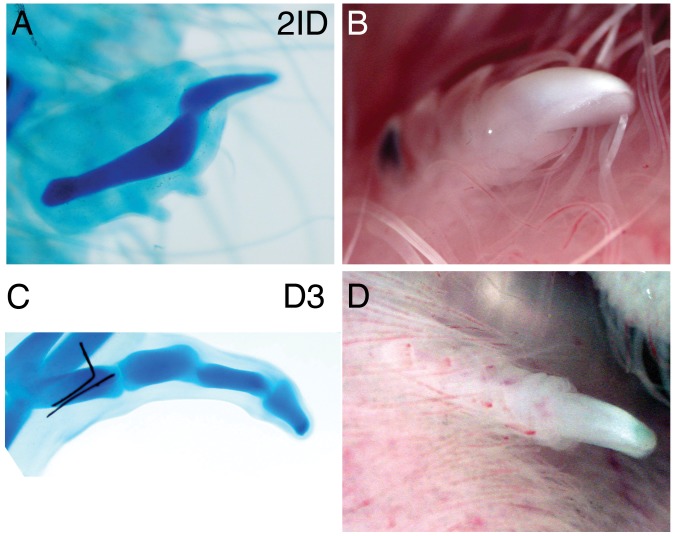
Bambi and Sp8 expression mark digit tips and their absence shows that chick wing digits 2 and 3 are truncated
- PMID: 23285181
- PMCID: PMC3532063
- DOI: 10.1371/journal.pone.0052781
Bambi and Sp8 expression mark digit tips and their absence shows that chick wing digits 2 and 3 are truncated
Abstract
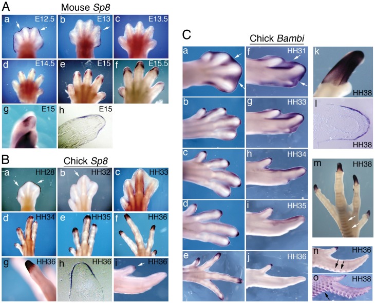
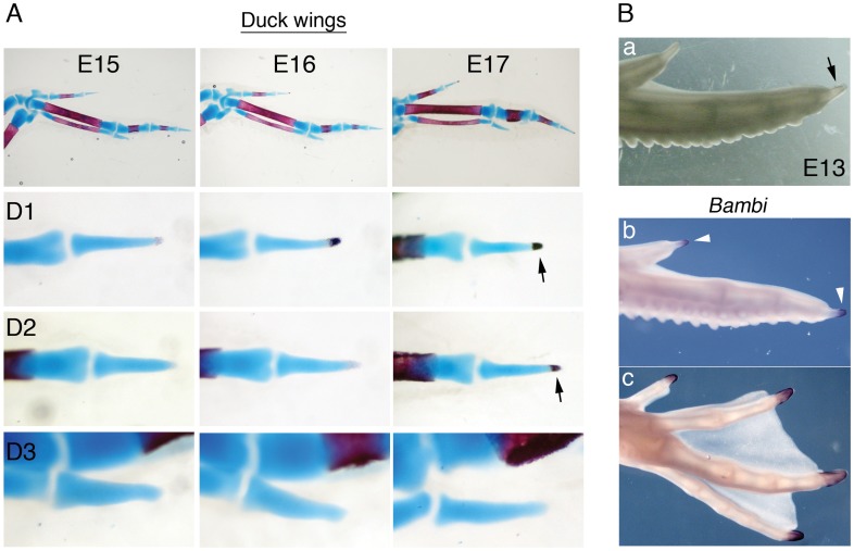
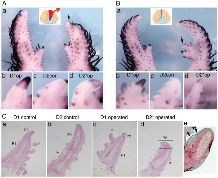
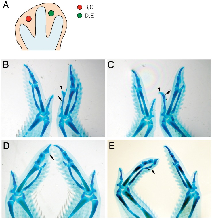
An often overlooked aspect of digit development is the special nature of the terminal phalanx, a specialized structure with characteristics distinct from other phalanges, for example the presence of ectodermal derivatives such as nails and claws. Here, we describe the unique ossification pattern of distal phalanges and characteristic gene expression in the digit tips of chick and duck embryos. Our results show that the distal phalanx of chick wing digit 1 is a genuine tip with a characteristic ossification pattern and expression of Bambi and Sp8; however, the terminal phalanx of digits 2* and 3 is not a genuine tip, and these are therefore truncated digits. Bambi and Sp8 expression in the chick wing provides a direct molecular assessment of digit identity changes after experimental manipulations of digit primordia. In contrast, digits 1 and 2 of the duck wing both possess true tips. Although chick wing-tip development was not rescued by application of Fgf8, this treatment induced the development of extra phalanges. Grafting experiments show that competence for tip formation, including nails, is latent in the interdigital tissue. Our results deepen understanding of the mechanisms of digit tip formation, highlighting its developmental autonomy and modular nature, with implications for digit reduction or loss during evolution. * Numbering of wing digits is 1, 2, 3 from anterior to posterior.
Conflict of interest statement
Figures

Similar articles
-
Comparative analysis of 3D expression patterns of transcription factor genes and digit fate maps in the developing chick wing.PLoS One. 2011 Apr 22;6(4):e18661. doi: 10.1371/journal.pone.0018661. PLoS One. 2011. PMID: 21526123 Free PMC article.
-
Transcriptomic analysis of avian digits reveals conserved and derived digit identities in birds.Nature. 2011 Sep 4;477(7366):583-6. doi: 10.1038/nature10391. Nature. 2011. PMID: 21892187
-
Inhibition of Shh signalling in the chick wing gives insights into digit patterning and evolution.Development. 2016 Oct 1;143(19):3514-3521. doi: 10.1242/dev.137398. Development. 2016. PMID: 27702785 Free PMC article.
-
The developmental evolution of avian digit homology: an update.Theory Biosci. 2005 Nov;124(2):165-83. doi: 10.1007/BF02814482. Epub 2005 Sep 12. Theory Biosci. 2005. PMID: 17046354 Review.
-
How is digit identity determined during limb development?Dev Growth Differ. 2013 Jan;55(1):130-8. doi: 10.1111/dgd.12022. Epub 2012 Dec 12. Dev Growth Differ. 2013. PMID: 23230964 Review.
Cited by
-
The receptor genes PfBMPR1B and PfBAMBI are involved in regulating shell biomineralization in the pearl oyster Pinctada fucata.Sci Rep. 2017 Aug 23;7(1):9219. doi: 10.1038/s41598-017-10011-y. Sci Rep. 2017. PMID: 28835628 Free PMC article.
-
Fgf signalling triggers an intrinsic mesodermal timer that determines the duration of limb patterning.Nat Commun. 2023 Sep 20;14(1):5841. doi: 10.1038/s41467-023-41457-6. Nat Commun. 2023. PMID: 37730682 Free PMC article.
-
Completion of neural crest cell production and emigration is regulated by retinoic-acid-dependent inhibition of BMP signaling.Elife. 2022 Apr 8;11:e72723. doi: 10.7554/eLife.72723. Elife. 2022. PMID: 35394423 Free PMC article.
-
Changes in Evolutionary Developmental Control Points in the Amniote Limb May Explain Hyperphalangy.Mol Biol Evol. 2025 Jun 4;42(6):msaf113. doi: 10.1093/molbev/msaf113. Mol Biol Evol. 2025. PMID: 40484714 Free PMC article.
-
Retinoic acid, an essential component of the roof plate organizer, promotes the spatiotemporal segregation of dorsal neural fates.Development. 2024 Oct 1;151(19):dev202973. doi: 10.1242/dev.202973. Epub 2024 Sep 30. Development. 2024. PMID: 39250350 Free PMC article.
References
-
- Zeller R, Lopez-Rios J, Zuniga A (2009) Vertebrate limb bud development: moving towards integrative analysis of organogenesis. Nat Rev Genet 10: 845–858. - PubMed
-
- Fernandez-Teran M, Ros MA (2008) The Apical Ectodermal Ridge: morphological aspects and signaling pathways. Int J Dev Biol 52: 857–871. - PubMed
-
- Kronenberg HM (2003) Developmental regulation of the growth plate. Nature 423: 332–336. - PubMed
-
- Dahn RD, Fallon JF (2000) Interdigital regulation of digit identity and homeotic transformation by modulated BMP signaling. Science 289: 438–441. - PubMed
-
- Sanz-Ezquerro JJ, Tickle C (2003) Fgf signaling controls the number of phalanges and tip formation in developing digits. Curr Biol 13: 1830–1836. - PubMed
Publication types
MeSH terms
Substances
LinkOut - more resources
Full Text Sources
