. 2012;7(12):e52886.
doi: 10.1371/journal.pone.0052886. Epub 2012 Dec 21.

Integrin Beta 1 suppresses multilayering of a simple epithelium

Affiliations

Integrin Beta 1 suppresses multilayering of a simple epithelium

Jichao Chen et al. PLoS One. 2012.

Abstract

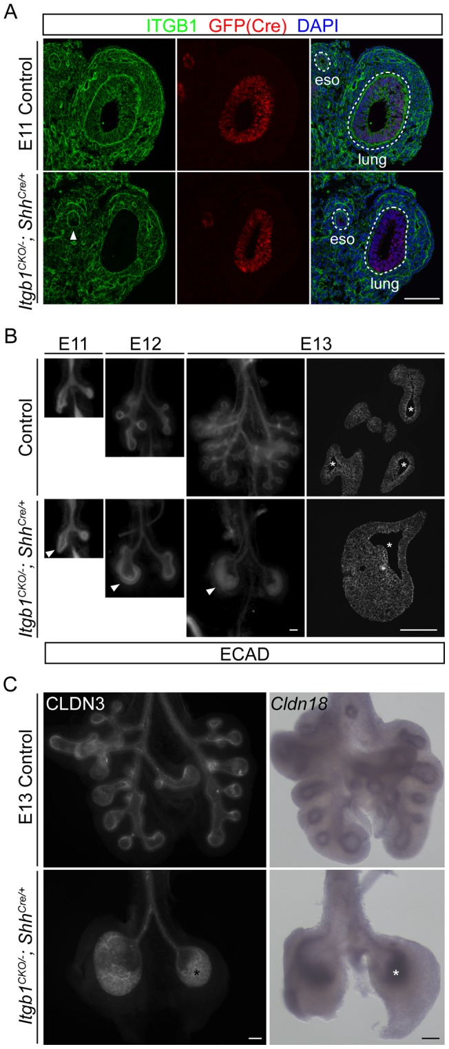
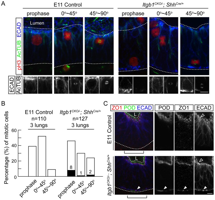
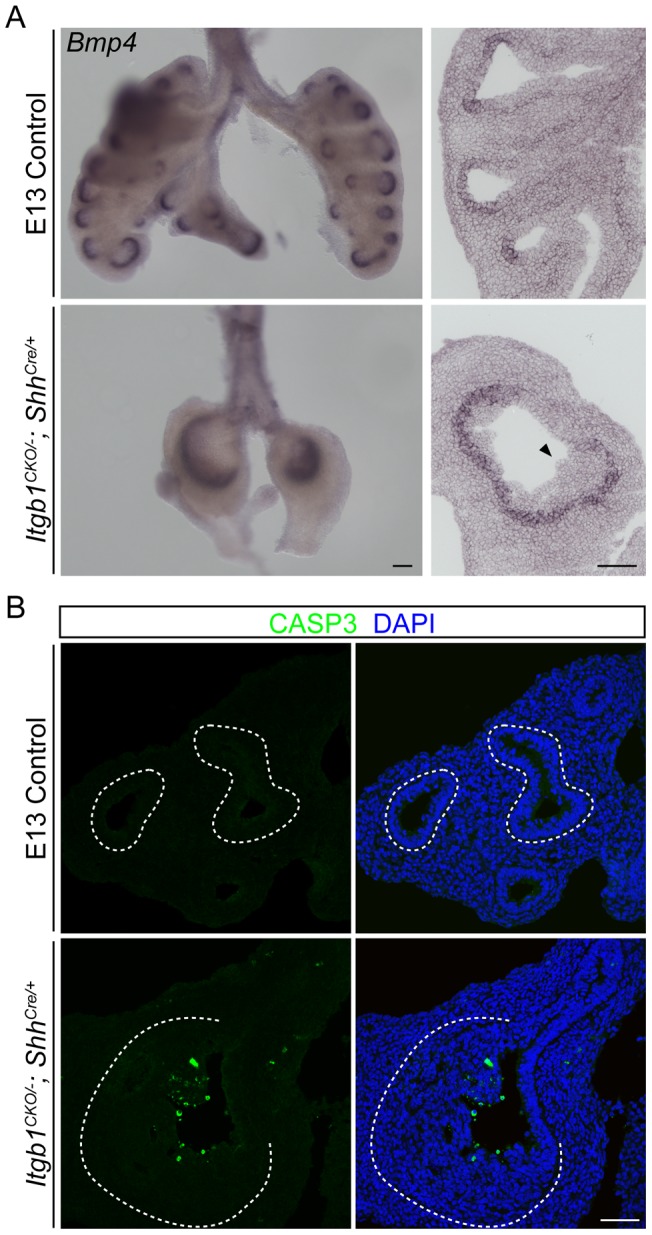
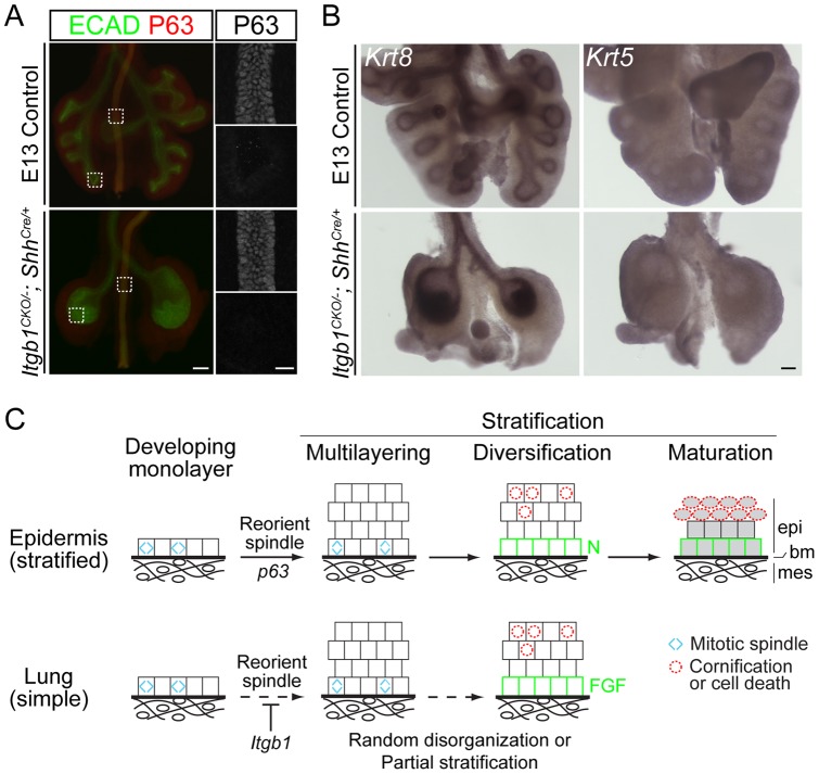
Epithelia are classified as either simple, a single cell layer thick, or stratified (multilayered). Stratified epithelia arise from simple epithelia during development, and transcription factor p63 functions as a key positive regulator of epidermal stratification. Here we show that deletion of integrin beta 1 (Itgb1) in the developing mouse airway epithelium abrogates airway branching and converts this monolayer epithelium into a multilayer epithelium with more than 10 extra layers. Mutant lung epithelial cells change mitotic spindle orientation to seed outer layers, and cells in different layers become molecularly and functionally distinct, hallmarks of normal stratification. However, mutant lung epithelial cells do not activate p63 and do not switch to the stratified keratin profile of epidermal cells. These data, together with previous data implicating Itgb1 in regulation of epidermal stratification, suggest that the simple-versus-stratified developmental decision may involve not only stratification inducers like p63 but suppressors like Itgb1 that prevent simple epithelia from inappropriately activating key steps in the stratification program.


Conflict of interest statement

Competing Interests: The authors have declared that no competing interests exist.

Figures

Figure 1
Figure 1. Epithelial inactivation of Itgb1 inhibits branching and leads to a multilayer epithelium during lung development.
(A) Section immunostaining showing complete loss of ITGB1 specifically in the lung epithelial cells, but not the surrounding mesenchymal cells as early as embryonic day 11 (E11) in the Itgb1CKO/−; ShhCre/+ mutant. Sections were co-stained for GFP to visualize cells expressing the GFP-CRE fusion protein under the control of the Shh promoter, and nuclei were counter-stained with 4′,6-diamidino-2-phenylindole (DAPI). The arrowhead indicates partial loss of ITGB1 in the ventral half of the esophagus epithelium in the Itgb1CKO/−; ShhCre/+ mutant. The dashed lines demarcate the basal side of the lung and esophagus (eso) epithelia. Scale bar, 100 um. (B) Whole-mount (left three columns) and section (right column) immunolocalization of E-Cadherin (ECAD) showing the inhibition of branching and progressive formation of a multilayer lung epithelium (arrowheads) at sequential embryonic days (E11, E12, E13) in the Itgb1CKO/−; ShhCre/+ mutant. Because of the three-dimensional structure of the lung, the epithelium can artificially appear multilayered in tangential sections through the epithelium. The number of epithelial layers can only be accurately assessed in regions of sections where the lumenal space (asterisks) is visible; note that there are more than 10 cell layers in the E13 mutant lung. Scale bar, 100 um. (C) Whole-mount immunostaining (left panels) and in situ hybridization (right panels) of E13 lungs showing that cells (asterisks) of the multilayer epithelium in the Itgb1CKO/−; ShhCre/+ mutant lung maintain expression of epithelial markers CLDN3 (left panels) and Cldn18 (right panels). Scale bar, 100 um.
Figure 2
Figure 2. Itgb1CKO/−; ShhCre/+ mutant lung epithelial cells reorient mitotic spindles and show partial loss of apical-basal polarity.
(A) Confocal sections of whole-mount E11 lungs immunostained for markers of mitosis (phospho-Histone 3, pH3), mitotic spindles (acetylated Tubulin, AcTUB) and the epithelium (E-Cadherin, ECAD). Representative images are shown for mitotic cells with no obvious mitotic spindle (prophase) and with the mitotic spindle orientated between 0 to 45 degrees (0°∼45°) or 45 to 90 degrees (45°∼90°) relative to the lumenal surface of the epithelium. Mitotic cells in the control lung are exclusively localized within one nuclear diameter of the lumenal surface of the epithelium. The Itgb1CKO/−; ShhCre/+ mutant lung contains mitotic cells localized away from the lumenal surface and sometimes on the basal surface of the epithelium. Grey scale images of the ECAD and AcTUB staining are shown below for the mitotic cells of interest. Note the intense ECAD puncta associated with the basally located Itgb1 mutant nuclei. The upper and lower dashed lines demarcate the lumenal (Lumen) and basal side of the epithelium, respectively. Scale bar, 10 um. (B) Quantification of the percentage of cells in each category in (A) out of 110 (control) and 127 (mutant) mitotic cells from 3 E11 lungs. The filled columns and the associated numbers indicate the percentage of mitotic cells that are located more than one nuclear diameter from the lumenal surface of the epithelium in the mutant. The control and mutant epithelia are significantly different in the distributions of mitotic cells between the 0°∼45°and 45°∼90° categories (52% and 9% in the control versus 30% and 24% in the mutant, p = 0.0004, Chi-square test), and in the number of mitotic nuclei located more than one nuclear diameter away from the lumenal surface of the epithelium (0 out of 110 in the control versus 14 out of 127 in the mutant, p = 0.0001, Fisher's exact test). (C) Confocal sections of whole-mount immunostained E11 lungs reveal basally located (arrowheads) markers of apical membrane (Podocalyxin-like, POD) and tight junction (ZO1) in the Itgb1CKO/−; ShhCre/+ mutant. In the control lung, cell junction protein E-Cadherin (ECAD) is localized between cells on the basal-lateral side of the epithelium and most concentrated as puncta (hollow arrowhead) close to the lumenal side. In the Itgb1CKO/−; ShhCre/+ mutant, additional ECAD puncta are localized to the basal side of the epithelium (arrowhead). These ectopic ECAD puncta are not obvious at low magnification or on sections (e.g., Figure 1B). The upper and lower dashed lines demarcate the lumenal (L) and basal side of the epithelium, respectively. Grey scale images are shown of the bracketed regions. Scale bar, 10 um.
Figure 3
Figure 3. Differences among cells in different layers of the multilayer Itgb1 mutant lung epithelium.
(A) Whole-mount (left column) and section (right column) in situ hybridization of E13 lungs showing that the expression of Bmp4 remains restricted to the distal branching epithelium in the Itgb1CKO/−; ShhCre/+ mutant, and that only cells on the basal side, but not on the lumenal side (arrowhead), of the multilayer epithelium in the Itgb1CKO/−; ShhCre/+ mutant lung express Bmp4. Scale bar, 100 um. (B) Section immunostaining of E13 lungs showing that cleaved Caspase-3 (CASP3), a marker of apoptosis, is preferentially expressed by cells located on the lumenal side of the multilayer epithelium in the Itgb1CKO/−; ShhCre/+ mutant lung. Nuclei were counter-stained with DAPI. The dashed lines demarcate the basal side of the lung epithelium. Scale bar, 50 um.
Figure 4
Figure 4. The multilayer epithelium in the Itgb1CKO/−; ShhCre/+ mutant lung expresses markers of simple epithelium.
(A) Stereoscope (left; scale bar, 100 um) and confocal (right; scale bar, 20 um) images of E13 whole-mount lungs immunostained for E-Cadherin (ECAD) and P63. The boxed regions in the left panels are imaged by confocal microscopy and shown in the right panels. P63 is expressed in the esophagus (top), but not in the branching regions of the lungs (bottom), of both control and Itgb1CKO/−; ShhCre/+ mutant embryos. (B) Whole-mount in situ hybridization shows that the epithelia of both control and Itgb1CKO/−; ShhCre/+ mutant lungs express a marker of simple epithelium (Krt8), but not a marker of stratified epithelium (Krt5) at E13. Scale bar, 100um. (C) A three-part model of epithelial stratification comparing epidermis and lung. Layer formation: regulation of mitotic spindle orientation (blue carets) drives formation of a multilayer epithelium; layer diversification: cell autonomous and/or non-autonomous signals lead to molecular and functional specialization of cells of different layers (green squares, cells with low Notch (N) pathway activity (epidermis) or high FGF pathway activity (lung); red dashed circles, cells undergoing cell death and cornification (epidermis) or cell death alone (lung)); layer maturation: activation of genes such as keratin genes specific for stratified epithelia and suppression of ones for simple epithelia leading to differentiation of the layers (shaded cells). Although the maturation step is shown last, the keratin profile switch in epidermis initiates in the monolayer and continues throughout stratification. In the absence of Itgb1, the normally simple lung epithelium may be in a random, disorganized state. However, the Itgb1 mutant epithelium shares two of the three features of epidermal stratification, suggesting that it undergoes partial stratification. epi, epithelium; bm, basement membrane; mes, mesenchyme.

Similar articles

Cited by

References

    1. Bragulla HH, Homberger DG (2009) Structure and functions of keratin proteins in simple, stratified, keratinized and cornified epithelia. J Anat 214: 516–559. - PMC - PubMed
    1. Lechler T, Fuchs E (2005) Asymmetric cell divisions promote stratification and differentiation of mammalian skin. Nature 437: 275–280. - PMC - PubMed
    1. Williams SE, Beronja S, Pasolli HA, Fuchs E (2011) Asymmetric cell divisions promote Notch-dependent epidermal differentiation. Nature 470: 353–358. - PMC - PubMed
    1. Ray S, Lechler T (2011) Regulation of asymmetric cell division in the epidermis. Cell Div 6: 12. - PMC - PubMed
    1. Koster MI, Roop DR (2007) Mechanisms regulating epithelial stratification. Annu Rev Cell Dev Biol 23: 93–113. - PubMed

Publication types

MeSH terms