Opposite role of tumor necrosis factor receptors in dextran sulfate sodium-induced colitis in mice
- PMID: 23285227
- PMCID: PMC3532169
- DOI: 10.1371/journal.pone.0052924
Opposite role of tumor necrosis factor receptors in dextran sulfate sodium-induced colitis in mice
Abstract
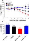
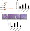
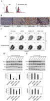
Tumor necrosis factor-α (TNF-α) is a key factor for the pathogenesis of inflammatory bowel diseases (IBD), whose function is known to be mediated by TNF receptor 1 (TNFR1) or 2. However, the precise role of the two receptors in IBD remains poorly understood. Herein, acute colitis was induced by dextran sulfate sodium (DSS) instillation in TNFR1 or 2-/- mice. TNFR1 ablation led to exacerbation of signs of colitis, including more weight loss, increased mortality, colon shortening and oedema, severe intestinal damage, and higher levels of myeloperoxidase compared to wild-type counterparts. While, TNFR2 deficiency had opposite effects. This discrepancy was reflected by alteration of proinflammatory cytokine and chemokine production in the colons. Importantly, TNFR1 ablation rendered enhanced apoptosis of colonic epithelial cells and TNFR2 deficiency conferred pro-apoptotic effects of lamina propria (LP)-immune cells, as shown by the decreased ratio of Bcl-2/Bax and enhanced nuclear factor (NF)-κB activity.
Conflict of interest statement
Figures


Similar articles
-
TNFR2 activates MLCK-dependent tight junction dysregulation to cause apoptosis-mediated barrier loss and experimental colitis.Gastroenterology. 2013 Aug;145(2):407-15. doi: 10.1053/j.gastro.2013.04.011. Epub 2013 Apr 22. Gastroenterology. 2013. PMID: 23619146 Free PMC article.
-
Protective role of tumor necrosis factor (TNF) receptors in chronic intestinal inflammation: TNFR1 ablation boosts systemic inflammatory response.Lab Invest. 2013 Sep;93(9):1024-35. doi: 10.1038/labinvest.2013.89. Epub 2013 Jul 29. Lab Invest. 2013. PMID: 23897411
-
Role of TNF receptors, TNFR1 and TNFR2, in dextran sodium sulfate-induced colitis.Inflamm Bowel Dis. 2009 Oct;15(10):1515-25. doi: 10.1002/ibd.20951. Inflamm Bowel Dis. 2009. PMID: 19479745
-
Epithelial Nuclear Factor-x03BA;B Activation in Inflammatory Bowel Diseases and Colitis-Associated Carcinogenesis.Digestion. 2016;93(1):40-6. doi: 10.1159/000441670. Epub 2016 Jan 14. Digestion. 2016. PMID: 26789263 Review.
-
Development, validation and implementation of an in vitro model for the study of metabolic and immune function in normal and inflamed human colonic epithelium.Dan Med J. 2015 Jan;62(1):B4973. Dan Med J. 2015. PMID: 25557335 Review.
Cited by
-
TNFR2 expression by CD4 effector T cells is required to induce full-fledged experimental colitis.Sci Rep. 2016 Sep 7;6:32834. doi: 10.1038/srep32834. Sci Rep. 2016. PMID: 27601345 Free PMC article.
-
MDSCs are involved in the protumorigenic potentials of GM-CSF in colitis-associated cancer.Int J Immunopathol Pharmacol. 2017 Jun;30(2):152-162. doi: 10.1177/0394632017711055. Epub 2017 May 23. Int J Immunopathol Pharmacol. 2017. PMID: 28534709 Free PMC article.
-
TNFR2 activates MLCK-dependent tight junction dysregulation to cause apoptosis-mediated barrier loss and experimental colitis.Gastroenterology. 2013 Aug;145(2):407-15. doi: 10.1053/j.gastro.2013.04.011. Epub 2013 Apr 22. Gastroenterology. 2013. PMID: 23619146 Free PMC article.
-
TNF blocking therapies and immunomonitoring in patients with inflammatory bowel disease.Mediators Inflamm. 2014;2014:172821. doi: 10.1155/2014/172821. Epub 2014 Mar 18. Mediators Inflamm. 2014. PMID: 24757282 Free PMC article. Review.
-
Redeeming an old foe: protective as well as pathophysiological roles for tumor necrosis factor in inflammatory bowel disease.Am J Physiol Gastrointest Liver Physiol. 2015 Feb 1;308(3):G161-70. doi: 10.1152/ajpgi.00142.2014. Epub 2014 Dec 4. Am J Physiol Gastrointest Liver Physiol. 2015. PMID: 25477373 Free PMC article. Review.
References
-
- Papadakis KA, Targan SR (2000) Role of cytokines in the pathogenesis of inflammatory bowel disease. Annu Rev Med 51: 289–298. - PubMed
-
- Kallias G, Douni E, Kassiotis G, Kontoyiannis D (1999) On the role of tumor necrosis factor and receptors in models of multiorgan failure, rheumatoid arthritis, multiple sclerosis and inflammatory bowel disease. Immuol Rev 169: 175–194. - PubMed
-
- MacEwan DJ (2002) TNF receptor subtype signaling: differences and cellular consequences. Cell Signal 14: 477–492. - PubMed
-
- Pimental-Munios FX, Seed B (1999) Regulated commitment of TNF signaling: a molecular switch for death or activation. Immunity 11: 783–793. - PubMed
Publication types
MeSH terms
Substances
LinkOut - more resources
Full Text Sources
Molecular Biology Databases
Research Materials

