Differential response of hippocampal subregions to stress and learning
- PMID: 23285257
- PMCID: PMC3532167
- DOI: 10.1371/journal.pone.0053126
Differential response of hippocampal subregions to stress and learning
Abstract
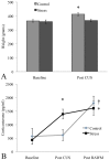
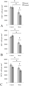
The hippocampus has two functionally distinct subregions-the dorsal portion, primarily associated with spatial navigation, and the ventral portion, primarily associated with anxiety. In a prior study of chronic unpredictable stress (CUS) in rodents, we found that it selectively enhanced cellular plasticity in the dorsal hippocampal subregion while negatively impacting it in the ventral. In the present study, we determined whether this adaptive plasticity in the dorsal subregion would confer CUS rats an advantage in a spatial task-the radial arm water maze (RAWM). RAWM exposure is both stressful and requires spatial navigation, and therefore places demands simultaneously upon both hippocampal subregions. Therefore, we used Western blotting to investigate differential expression of plasticity-associated proteins (brain derived neurotrophic factor [BDNF], proBDNF and postsynaptic density-95 [PSD-95]) in the dorsal and ventral subregions following RAWM exposure. Lastly, we used unbiased stereology to compare the effects of CUS on proliferation, survival and neuronal differentiation of cells in the dorsal and ventral hippocampal subregions. We found that CUS and exposure to the RAWM both increased corticosterone, indicating that both are stressful; nevertheless, CUS animals had significantly better long-term spatial memory. We also observed a subregion-specific pattern of protein expression following RAWM, with proBDNF increased in the dorsal and decreased in the ventral subregion, while PSD-95 was selectively upregulated in the ventral. Finally, consistent with our previous study, we found that CUS most negatively affected neurogenesis in the ventral (compared to the dorsal) subregion. Taken together, our data support a dual role for the hippocampus in stressful experiences, with the more resilient dorsal portion undergoing adaptive plasticity (perhaps to facilitate escape from or neutralization of the stressor), and the ventral portion involved in affective responses.
Conflict of interest statement
Figures


Similar articles
-
Region-specific response of the hippocampus to chronic unpredictable stress.Hippocampus. 2012 Jun;22(6):1338-49. doi: 10.1002/hipo.20970. Epub 2011 Jul 29. Hippocampus. 2012. PMID: 21805528
-
Emotionality modulates the impact of chronic stress on memory and neurogenesis in birds.Sci Rep. 2020 Sep 3;10(1):14620. doi: 10.1038/s41598-020-71680-w. Sci Rep. 2020. PMID: 32884096 Free PMC article.
-
Incidental (unreinforced) and reinforced spatial learning in rats with ventral and dorsal lesions of the hippocampus.Behav Brain Res. 2009 Aug 24;202(1):64-70. doi: 10.1016/j.bbr.2009.03.016. Epub 2009 Mar 21. Behav Brain Res. 2009. PMID: 19447282
-
[Effects of stress factors on adult hippocampus: molecular, cellular mechanisms and dorso-ventral gradient].Ross Fiziol Zh Im I M Sechenova. 2013 Jan;99(1):3-16. Ross Fiziol Zh Im I M Sechenova. 2013. PMID: 23659052 Review. Russian.
-
Neurogenesis along the septo-temporal axis of the hippocampus: are depression and the action of antidepressants region-specific?Neuroscience. 2013 Nov 12;252:234-52. doi: 10.1016/j.neuroscience.2013.08.017. Epub 2013 Aug 20. Neuroscience. 2013. PMID: 23973415 Review.
Cited by
-
Chronic unpredictable stress during adolescence protects against adult traumatic brain injury-induced affective and cognitive deficits.Brain Res. 2021 Sep 15;1767:147544. doi: 10.1016/j.brainres.2021.147544. Epub 2021 Jun 4. Brain Res. 2021. PMID: 34090883 Free PMC article.
-
Children's family income is associated with cognitive function and volume of anterior not posterior hippocampus.Nat Commun. 2020 Aug 12;11(1):4040. doi: 10.1038/s41467-020-17854-6. Nat Commun. 2020. PMID: 32788583 Free PMC article.
-
Medial Temporal Lobe Subregional Atrophy in Aging and Alzheimer's Disease: A Longitudinal Study.Front Aging Neurosci. 2021 Oct 15;13:750154. doi: 10.3389/fnagi.2021.750154. eCollection 2021. Front Aging Neurosci. 2021. PMID: 34720998 Free PMC article.
-
Altered hippocampal microstructure and function in children who experienced Hurricane Irma.Dev Psychobiol. 2021 Jul;63(5):864-877. doi: 10.1002/dev.22071. Epub 2020 Dec 16. Dev Psychobiol. 2021. PMID: 33325561 Free PMC article.
-
Polygenic risk for depression and anterior and posterior hippocampal volume in children and adolescents.J Affect Disord. 2024 Jan 1;344:619-627. doi: 10.1016/j.jad.2023.10.068. Epub 2023 Oct 18. J Affect Disord. 2024. PMID: 37858734 Free PMC article.
References
-
- Moser MB, Moser EI (1998) Functional differentiation in the hippocampus. Hippocampus 8: 608–619. - PubMed
-
- Maurer AP, Vanrhoads SR, Sutherland GR, Lipa P, McNaughton BL (2005) Self-motion and the origin of differential spatial scaling along the septo-temporal axis of the hippocampus. Hippocampus 15: 841–852. - PubMed
-
- Bannerman DM, Grubb M, Deacon RM, Yee BK, Feldon J, et al. (2003) Ventral hippocampal lesions affect anxiety but not spatial learning. Behav Brain Res 139: 197–213. - PubMed
-
- Bannerman DM, Rawlins JN, McHugh SB, Deacon RM, Yee BK, et al. (2004) Regional dissociations within the hippocampus–memory and anxiety. Neurosci Biobehav Rev 28: 273–283. - PubMed
Publication types
MeSH terms
Grants and funding
LinkOut - more resources
Full Text Sources
Medical

