. 2012;7(12):e53195.
doi: 10.1371/journal.pone.0053195. Epub 2012 Dec 28.

Striatal cholinergic interneurons display activity-related phosphorylation of ribosomal protein S6

Affiliations

Striatal cholinergic interneurons display activity-related phosphorylation of ribosomal protein S6

Jesus Bertran-Gonzalez et al. PLoS One. 2012.

Abstract

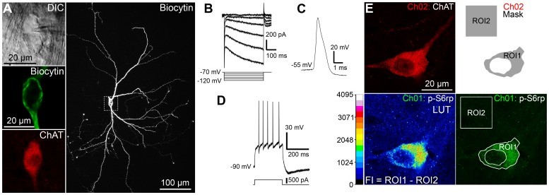
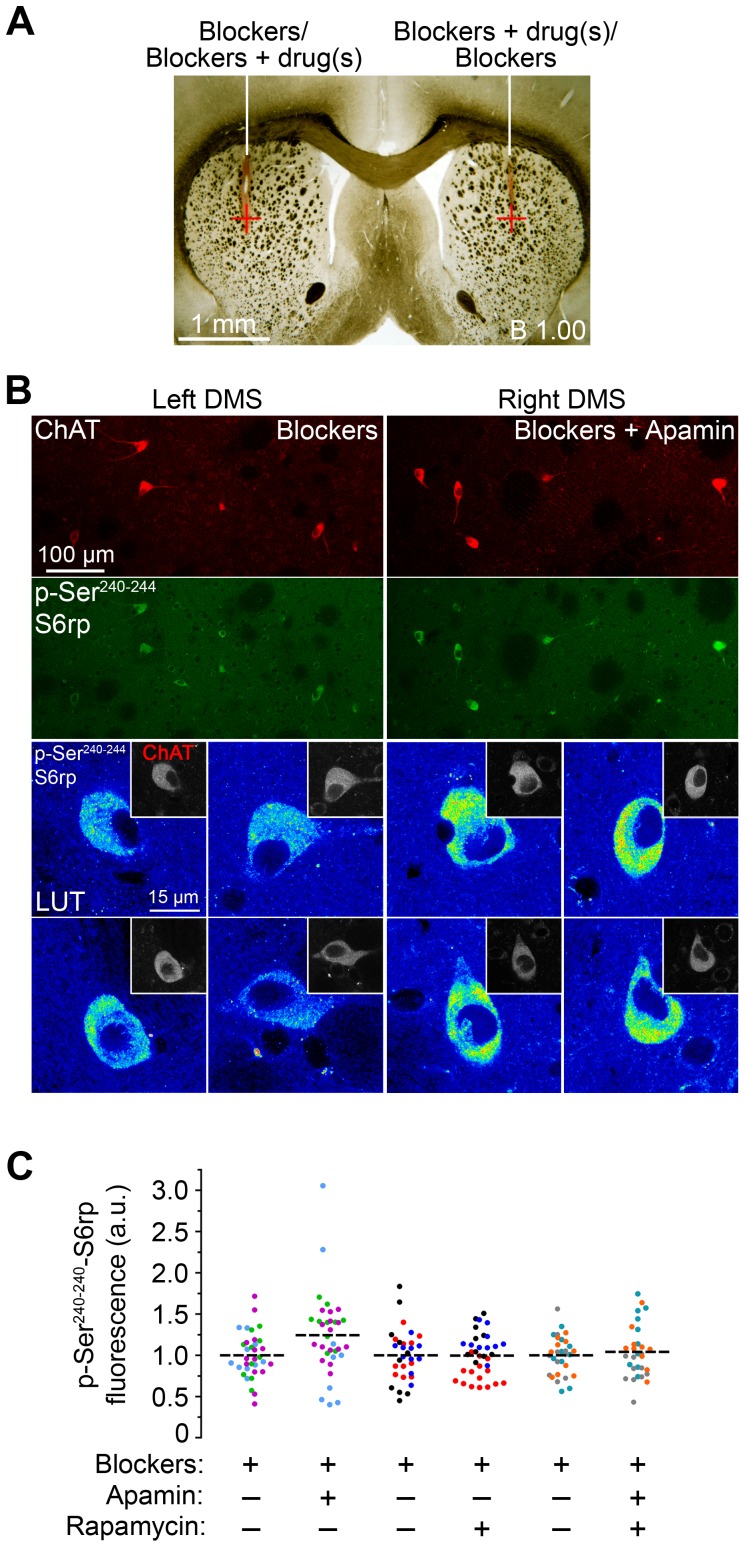
Cholinergic interneurons (CINs) provide the main source of acetylcholine to all striatal regions, and strongly modulate dopaminergic actions through complex regulation of pre- and post-synaptic acetylcholine receptors. Although striatal CINs have a well-defined electrophysiological profile, their biochemical properties are poorly understood, likely due to their low proportion within the striatum (2-3%). We report a strong and sustained phosphorylation of ribosomal protein S6 on its serine 240 and 244 residues (p-Ser²⁴⁰⁻²⁴⁴-S6rp), a protein integrant of the ribosomal machinery related to the mammalian target of the rapamycin complex 1 (mTORC1) pathway, which we found to be principally expressed in striatal CINs in basal conditions. We explored the functional relevance of this cellular event by pharmacologically inducing various sustained physiological activity states in CINs and assessing the effect on the levels of S6rp phosphorylation. Cell-attached electrophysiological recordings from CINs in a striatal slice preparation showed an inhibitory effect of tetrodotoxin (TTX) on action potential firing paralleled by a decrease in the p-Ser²⁴⁰⁻²⁴⁴-S6rp signal as detected by immunofluorescence after prolonged incubation. On the other hand, elevation in extracellular potassium concentration and the addition of apamin generated an increased firing rate and a burst-firing activity in CINs, respectively, and both stimulatory conditions significantly increased Ser²⁴⁰⁻²⁴⁴-S6rp phosphorylation above basal levels when incubated for one hour. Apamin generated a particularly large increase in phosphorylation that was sensitive to rapamycin. Taken together, our results demonstrate for the first time a link between the state of neuronal activity and a biochemical signaling event in striatal CINs, and suggest that immunofluorescence can be used to estimate the cellular activity of CINs under different pharmacological and/or behavioral conditions.


Conflict of interest statement

Competing Interests: The authors have declared that no competing interests exist.

Figures

Figure 1
Figure 1. Cholinergic interneurons (CINs) in the striatum show selective phosphorylation of S6rp in basal conditions.
(A) Low magnification image from a transcardially-fixed naïve rat brain section showing the striatal level analyzed in the present study. Inset corresponds to magnified right panels: low magnification images of the dorsal striatum double-stained with choline acetyltransferase (ChAT, green) and phosphorylated ribosomal protein S6 at Serine 240 and 244 residues (p-S6rp, red) showing substantial co-localization. Insets (bottom-left) are high magnification confocal images showing ChAT and p-S6rp immunoreactivity in two adjacent CINs. (B, C) Confocal sections of rat striatal tissue stained for two different pairs of phospho-serine residues at the C terminus of S6rp (B, Ser240–244 and C, Ser235–236, green) combined with ChAT (red) and DARPP-32 (D-32, magenta). Arrows indicate ChAT-immunoreactive neurons. (B, C; bottom right graphs) Intensity fluorescence study of p-Ser240–244 (B) and p-Ser235–236 (C) S6rp signal contained in MSNs (D-32 immunoreactive) and CINs (ChAT immunoreactive) in triple-stained sections. Data are mean ± SEM; n = 2 rats; 40 MSNs and 13 CINs quantified per group.
Figure 2
Figure 2. Cellular physiology and S6rp phosphorylation in CINs from viable striatal slices.
(A) A representative striatal ChAT-immunoreactive neuron labeled with biocytin during electrophysiological recording. Visualization of the neuron after difference interference contrast (DIC) illumination (top left A), post hoc single optical scan of biocytin (middle left A) and ChAT labeling (bottom left A), and confocal Z stack projection of biocytin labeling at low magnification (20×, right A). (B–D) Cellular physiological characteristics of the neuron in A under whole-cell patch-clamp. Current-voltage relationship recorded by stepping the cell to various hyperpolarizing membrane potentials (B). Under current-clamp configuration, whole-cell action potential (C) and depolarization-triggered action potential firing (D) were routinely sampled for comparisons with known CIN cellular characteristics. (E) Quantification of p-S6rp in a ChAT-immunoreactive neuron from a viable striatal slice after overnight fixation and immunofluorescence. The neuron was identified under the microscope and a high magnification optical section was taken sequentially (Ch01: p-Ser240–244S6rp; Ch02: ChAT). Fluorescence intensity (FI) of the phospho-S6rp signal in each neuron was studied by defining an image mask in Ch02 (ROI1: somatic area; ROI2: background in 15 um2 in the vicinity of the soma) and superimposing it onto Ch01. The mean gray value of the pixels contained in ROI1 and ROI2 was obtained for Ch01, and FI was defined as ROI1 minus ROI2. A 16 pseudo-color palette (Lookup Table, LUT) applied in Ch01 highlights the intensity of p-S6rp fluorescence. The LUT scale defines 16-color intervals according to pixel gray values in a 16-bit image.
Figure 3
Figure 3. Tetrodotoxin (TTX) inhibits basal cellular activity and reduces S6rp phosphorylation in striatal CINs.
(A) Cell-attached recording of a striatal cholinergic interneuron depicting TTX (100 nM) inhibition of spontaneous action potential firing. The open bar indicates time of TTX application. The bottom traces show an expanded time scale (n = 3 neurons). (B) High-magnification confocal images of 5 different striatal CINs (ChAT-immunoreactive, insets) and their corresponding p-Ser240–244-S6rp levels in slices incubated in physiological saline (vehicle) or plus TTX (1 µM). A 16 pseudo-color palette LUT highlights intensity of p-S6rp fluorescence. (C) Quantification of the p-S6rp signal in each striatal ChAT immunoreactive neuron after 1-hour incubation in control or TTX (1 µM). P-S6rp signal intensity for each neuron was calculated as in Figure 2E. In scatterplot, each dot corresponds to one neuron; each color corresponds to a different animal; dashed lines indicate the mean. Fluorescence values are normalized in arbitrary units (a.u.). Data were analyzed using unpaired Student t-test: p<0.0001; 81–88 quantified neurons per condition in 3 rats.
Figure 4
Figure 4. Different forms of cellular activity stimulation increase S6rp phosphorylation over basal levels in striatal CINs.
(A) Cell-attached recording of a striatal CIN showing a strong increase in action potential firing after elevation of extracellular potassium concentration (High K+, from 2.5 mM in pre-condition to 11.5 mM in post-condition; n = 3 neurons). Gray shade indicates time of elevated K+ application. Bottom traces show an expanded time scale in pre- and post- K+ elevation. (B) Cell-attached recording of a striatal CIN showing typical burst-firing behavior after application of apamin (100 nM; n = 7 neurons). Gray bar indicates time of apamin application. Bottom traces show an expanded time scale in pre- and post- apamin conditions. (C) High-magnification confocal images of 5 different striatal CINs (ChAT-immunoreactive, insets) and their corresponding p-Ser240–244-S6rp levels in slices incubated in physiological saline (vehicle), high K+ (elevated extracellular K+ to 11.5 mM) or apamin (100 nM). A 16 pseudo-color palette LUT highlights intensity of p-S6rp fluorescence. (D) Quantification of p-S6rp signal in striatal ChAT immunoreactive neurons in each incubation condition. The p-S6rp signal intensity for each neuron was calculated as in Figure 2E. (E) Cell-attached recording of a CIN in the presence of synaptic blockers picrotoxin (Pic, 100 µM), CNQX (10 µM) and DL-AP5 (AP5, 100 µM). Application of apamin (100 nM) induced burst-firing responses as in A (n = 9 neurons). (F) Quantification of the p-S6rp signal in striatal ChAT immunoreactive neurons in each incubation condition. Application of synaptic blockers (Block) did not alter baseline p-S6rp signal nor did it inhibit the stimulatory effects of apamin (100 nM). In scatterplots (D and F), each dot corresponds to one neuron; each color corresponds to a different animal; dashed lines indicate the mean. Fluorescence values are normalized in arbitrary units (a.u.). Data were analyzed with one-way ANOVA: D: Effect of drug: F(2,247) = 20.11, p<0.0001; Bonferroni post hoc: Vehicle vs. High K+: p<0.001; Vehicle vs. Apamin: p<0.001; High K+ vs. Apamin: p<0.05; 81–87 neurons quantified per condition in 4 rats. F: Effect of drug: F(2,201) = 9.503, p<0.0001; Bonferroni post hoc: Vehicle vs. Block+Apamin: p<0.001; Block vs. Block+Apamin: p<0.01; 66–70 quantified neurons per condition in 5 rats).
Figure 5
Figure 5. Rapamycin prevents apamin-stimulated S6rp phosphorylation but not cellular activity in striatal CINs.
(A) Cell-attached recording of a CIN in the presence of synaptic blockers picrotoxin (Pic, 100 µM), CNQX (10 µM) and DL-AP5 (AP5, 100 µM) showing changes in firing after application of rapamycin (dark gray) and apamin (black). As shown in the bottom traces (n = 3 neurons), application of rapamycin (Rapa, 1 µM) did not alter spontaneous action potential firing nor did it affect the burst-firing response induced by final apamin application (Apa, 100 nM, n = 3 neurons). (B) High-magnification confocal images of representative striatal CINs (ChAT-immunoreactive, insets) and their corresponding p-Ser240–244-S6rp levels in slices incubated in the presence of synaptic blockers (Bl), plus 100 nM apamin (Bl+Apa) and plus 1 µM rapamycin (Bl+Apa+Rapa). (C) Quantification of the p-S6rp signal in striatal ChAT immunoreactive neurons in each incubation condition (intensity for each neuron was calculated as in Figure 2E). A 16 pseudo-color palette LUT highlights the intensity of p-S6rp fluorescence. In scatterplot, each dot corresponds to one neuron; each color corresponds to a different animal; dashed lines indicate the mean. Fluorescence values are normalized into arbitrary units (a.u.). Data were analyzed with one-way ANOVA (Effect of drug: F(2,132) = 15.54, p<0.0001; Bonferroni post hoc: Blockers vs. Blockers+Apamin; p<0.001; Blockers+Apamin vs. Blockers+Apamin+Rapamycin; p<0.001; 45 quantified neurons per condition in 3 rats).
Figure 6
Figure 6. Effects of apamin and rapamycin on S6rp phosphorylation in CINs are reproduced in vivo.
(A) Low magnification image of a rat brain section showing the targeted striatal region bilaterally (red cross). Rats received either synaptic blocker solution (blockers; picrotoxin, 150 µM; CNQX disodium, 1 mM; DL-AP5, 1 mM) or the same solution plus experimental drug(s) (apamin, 100 nM and/or rapamycin, 1 µM) in contralateral hemispheres. (B) Confocal images from a transcardially-fixed rat brain previously injected with blockers (left-side dorsal striatum, Left DMS) and blockers plus apamin (right-side dorsal striatum, Right DMS) showing double staining for ChAT and p-Ser240–244-S6rp. Top panels are low-magnification images showing several cholinergic interneurons in the same focal plane. Bottom panels are higher magnification images showing p-S6rp signal intensity in CINs from the left (blockers) and right (blockers + apamin) striata of the same animal. Insets show corresponding ChAT staining. (C) Quantification of p-S6rp signal in striatal ChAT immunoreactive neurons after each combination of injections (intensity for each neuron was calculated as in Figure 2E). A 16 pseudo-color palette LUT highlights the intensity of p-S6rp fluorescence. In the scatterplot, each dot corresponds to one neuron; each color corresponds to a different animal; dashed lines indicate the mean. Fluorescence values are normalized into arbitrary units (a.u.). Data were analyzed with one-way ANOVA (Effect of drug: F(5,188) = 2.932, p = 0.0142, Bonferroni post hoc: Blockers vs. Blockers+Apamin; p<0.01; 30–35 quantified neurons per condition in 9 rats).

References

    1. Alexander GE, DeLong MR, Strick PL (1986) Parallel organization of functionally segregated circuits linking basal ganglia and cortex. Annual review of neuroscience 9: 357–381. - PubMed
    1. Gerfen CR, Surmeier DJ (2011) Modulation of striatal projection systems by dopamine. Annual review of neuroscience 34: 441–466. - PMC - PubMed
    1. Kawaguchi Y, Wilson CJ, Augood SJ, Emson PC (1995) Striatal interneurones: chemical, physiological and morphological characterization. Trends in neurosciences 18: 527–535. - PubMed
    1. Tepper JM, Bolam JP (2004) Functional diversity and specificity of neostriatal interneurons. Current opinion in neurobiology 14: 685–692. - PubMed
    1. Bolam JP (1984) Synapses of identified neurons in the neostriatum. Ciba Foundation symposium 107: 30–47. - PubMed

Publication types

Substances