. 2012;7(12):e53319.
doi: 10.1371/journal.pone.0053319. Epub 2012 Dec 28.

cGMP-dependent protein kinase contributes to hydrogen sulfide-stimulated vasorelaxation

Affiliations

cGMP-dependent protein kinase contributes to hydrogen sulfide-stimulated vasorelaxation

Mariarosaria Bucci et al. PLoS One. 2012.

Abstract

A growing body of evidence suggests that hydrogen sulfide (H₂S) is a signaling molecule in mammalian cells. In the cardiovascular system, H₂S enhances vasodilation and angiogenesis. H₂S-induced vasodilation is hypothesized to occur through ATP-sensitive potassium channels (K(ATP)); however, we recently demonstrated that it also increases cGMP levels in tissues. Herein, we studied the involvement of cGMP-dependent protein kinase-I in H₂S-induced vasorelaxation. The effect of H₂S on vessel tone was studied in phenylephrine-contracted aortic rings with or without endothelium. cGMP levels were determined in cultured cells or isolated vessel by enzyme immunoassay. Pretreatment of aortic rings with sildenafil attenuated NaHS-induced relaxation, confirming previous findings that H₂S is a phosphodiesterase inhibitor. In addition, vascular tissue levels of cGMP in cystathionine gamma lyase knockouts were lower than those in wild-type control mice. Treatment of aortic rings with NaHS, a fast releasing H₂S donor, enhanced phosphorylation of vasodilator-stimulated phosphoprotein in a time-dependent manner, suggesting that cGMP-dependent protein kinase (PKG) is activated after exposure to H₂S. Incubation of aortic rings with a PKG-I inhibitor (DT-2) attenuated NaHS-stimulated relaxation. Interestingly, vasodilatory responses to a slowly releasing H₂S donor (GYY 4137) were unaffected by DT-2, suggesting that this donor dilates mouse aorta through PKG-independent pathways. Dilatory responses to NaHS and L-cysteine (a substrate for H₂S production) were reduced in vessels of PKG-I knockout mice (PKG-I⁻/⁻). Moreover, glibenclamide inhibited NaHS-induced vasorelaxation in vessels from wild-type animals, but not PKG-I⁻/⁻, suggesting that there is a cross-talk between K(ATP) and PKG. Our results confirm the role of cGMP in the vascular responses to NaHS and demonstrate that genetic deletion of PKG-I attenuates NaHS and L-cysteine-stimulated vasodilation.


Conflict of interest statement

Competing Interests: The authors have declared that no competing interests exist.

Figures

Figure 1
Figure 1. The PDE5 inhibitor sildenafil differentially affects NO and H2S-regulated vascular tone.
(A) Incubation of isolated aortic rings with sildenafil (1 nM) significantly inhibited NaHS-induced vasodilatation. (B) Incubation of isolated aortic rings with sildenafil (1 nM) significantly enhanced SNAP-induced vasodilatation. *** p<0.001 vs vehicle (dH2O), n = 6 for each group.
Figure 2
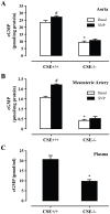
Figure 2. CSE deficiency reduces cGMP levels. cGMP levels in the aorta (A), mesenteric artery (B) and plasma (C) of CSE−/− mice were significantly lower than those from CSE+/+ mice.
Sodium nitroprusside (SNP, 10 µM) significantly increased cGMP levels in aorta (B) and mesenteric artery (C) from CSE+/+ mice but not CSE−/− mice; * p<0.05 vs CSE+/+ mice, #p<0.05 basal, n = 5 for each group.
Figure 3
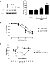
Figure 3. H2S activates PKG and triggers vasodilatation.
(A) Mouse aorta was incubated with NaHS (50 µM) for the indicated time and VASP phosphorylation on Ser239 was determined. Left: representative blot; right: quantitation of scanned autoradiograms, *p<0.05 vs vehicle, n = 3. (B) Incubation of aortic rings with the selective inhibitor of PKG, DT-2 (1, 3 µM) significantly inhibited NaHS-induced vasodilatation. TAT peptide (3 µM) was used as a control; *** p<0.001 vs. vehicle (dH2O), n = 6 for each group. (C) Mice were injected with vehicle or DT-2 (100 nmoles, ip); after 15 min NaHS (1 µmol/kg) was administered subcutaneously. Systolic blood pressure (SBP) was monitored in conscious mice; *** p<0.001 vs vehicle, n = 4 for each group.
Figure 4
Figure 4. GYY4137-induced relaxation is independent of PKG.
Aortic smooth muscle cells were exposed to the indicated concentration of NaHS (A) or GYY4137 (B) and cGMP levels were determined after 5 min. *p<0.05 control, n = 4 for each group. (C) Incubation of isolated aortic rings with PKG selective inhibitor DT-2 (3 µM) did not affect GYY4137-induced vasodilatation; n = 6 for each group.
Figure 5
Figure 5. PKG contributes to the relaxing effect of exogenous and endogenous H2S.
(A) Mouse aortas from wild-type or PKG-I−/− animals were pre-treated with vehicle or glibenclamide (10 µM, 30 min) and then incubated with the indicated concentration of NaHS (n = 6 rings harvested from 3–4 animals); dashed lines are used for wild-type animals, while solid lines are used for knockouts. (B) L-cysteine-induced vasodilatation of aortic rings pre-contracted with phenylephrine from wild-type and PKG-I−/− mice. Note that cumulative concentration-response curves to L-cysteine were significantly different among the different strains of mice used CD1 vs 129/Sv (WT); °°° p<0.001 vs. CD1, *** p<0.001 vs WT, n = 8 rings harvested from 3–4 animals for each group (C) Representative blot and quantitation depicting aortic CSE expression in 129/Sv vs CD-1; n = 3 for each group, *p<0.05. (D) Representative blot showing expression of CSE in aortic homogenates in wild-type and PKG-I−/− mice. Experiments were performed twice with similar results.
Figure 6
Figure 6. Role of endothelium in H2S induced-vasodilatation.
(A) L-cysteine-induced vasodilatation was significantly impaired in aortic rings without endothelium (–end). (B) NaHS-induced vasodilatation is not affected by endothelium removal; *** p<0.001 vs –end, n = 6 for each group. (C) Representative photomicrographs of aortas stained with a CSE antibody and counter-stained for von-Willebrant factor, smooth muscle α-actin (SMA) and DAPI, showing localization of CSE.

References

    1. Mustafa A, Gadalla M, Snyder S (2009) Signaling by Gasotransmitters. Sci Signal 2: re2. - PMC - PubMed
    1. Wang R (2002) Two's company, three's a crowd – Can H2S be the third endogenous gaseous transmitter? FASEB J 16: 1792–1798. - PubMed
    1. Sessa W (2004) eNOS at a glance. J Cell Sci 117: 2427–2429. - PubMed
    1. Motterlini R, Otterbein L (2010) The therapeutic potential of carbon monoxide. Nat Rev Drug Discov 9: 728–743. - PubMed
    1. Szabó C (2007) Hydrogen sulphide and its therapeutic potential. Nat Rev Drug Discov 6: 917–935. - PubMed

Publication types

MeSH terms

Substances

LinkOut - more resources