. 2012;7(12):e53817.
doi: 10.1371/journal.pone.0053817. Epub 2012 Dec 20.

Alveolar type II cells possess the capability of initiating lung tumor development

Affiliations

Alveolar type II cells possess the capability of initiating lung tumor development

Chuwen Lin et al. PLoS One. 2012.

Abstract

Identifying cells of tumor origin is a fundamental question in tumor biology. Answers to this central question will not only advance our understanding of tumor initiation and progression but also have important therapeutic implications. In this study, we aimed to uncover the cells of origin of lung adenocarcinoma, a major subtype of non-small cell lung cancer. To this end, we developed new mouse models of lung adenocarcinoma that enabled selective manipulation of gene activity in surfactant associated protein C (SPC)-expressing cells, including alveolar type II cells and bronchioalveolar stem cells (BASCs) that reside at the bronchioalveolar duct junction (BADJ). Our findings showed that activation of oncogenic Kras alone or in combination with the removal of the tumor suppressor p53 in SPC⁺ cells resulted in development of alveolar tumors. Similarly, sustained EGF signaling in SPC⁺ cells led to alveolar tumors. By contrast, BASCs failed to proliferate or produce tumors under these conditions. Importantly, in a mouse strain in which Kras/p53 activity was selectively altered in type II cells but not BASCs, alveolar tumors developed while BADJs retained normal architecture. These results confirm and extend previous findings and support a model in which lung adenocarcinoma can initiate in alveolar type II cells. Our results establish the foundation for elucidating the molecular mechanisms by which lung cancer initiates and progresses in a specific lung cell type.


Conflict of interest statement

Competing Interests: The authors have declared that no competing interests exist.

Figures

Figure 1
Figure 1. SPCCreER and SPCrtTA mice allow manipulation of gene activities in alveolar type II cells.
(A) Schematic diagram depicting gene targeting of a CreERT2tPA cassette at the translational start (ATG) of the mouse SPC genomic locus (exons are numbered). tPA contains three copies of the polyadenylation signal (PA). The resulting allele is designated SPCCreER in this study. Insertion of CreER disrupts the production of SPC mRNA but heterozygous SPCCreER mice are viable and exhibit no obvious phenotypes, similar to previous reports . (B) Schematic diagram illustrating the introduction of an rtTAtPA cassette at the translational start (ATG) of the mouse SPC genomic locus contained within a BAC. The engineered BAC was subsequently modified to introduce a tetO-Cre cassette at the translational start (ATG) of the Nudt18 genomic locus located approximately 57 kb upstream of the translational start of SPC. The transgenic mouse line carrying the modified BAC is designated SPCrtTA in this study. (C) Immunostaining and immunohistochemistry to assess the specificity and efficiency of SPCCreER and SPCrtTA mouse lines. (D–F) Immunostaining of lung sections from adult SPCCreER/+; ROSA26mTmG/+ mice. Without tamoxifen (TM) injection, low levels of CreER activity in alveolar type II cells (identified by anti-SPC) resulted in eGFP expression (detected by anti-GFP antibodies) from the ROSA26mTmG allele by removing sequences that block its expression. The levels of leaky CreER expression in alveolar type II cells seem to vary from animal to animal (D, E) and could even differ between lung lobes of a single uninjected animal. TM administration led to a dramatic increase in the number of eGFP-labeled type II cells (F), indicating efficient activation of CreER by TM. In addition to type II cells, SPC+CC10+ bronchioalveolar stem cells (BASCs) located at the bronchioalveolar duct junction (BADJ) were also efficiently labeled by eGFP upon TM injection (I). The insets in (I) show individual images of eGFP/SPC/CC10 immunostaining of the same BASC (arrow) or eGFP/SPC immunostaining of the same type II cell (arrow). The percentage of distinct cell types labeled by eGFP upon TM administration is summarized in (J). At least five mice and more than three sections per mouse were examined. Since BASCs are less prevalent than type II, type I and Clara cells, all BASCs present on a given section were counted. The proportion of lineage-labeled cells was scored as follows: type II cell (ratio of SPC+eGFP+/eGFP+); BASC (ratio of SPC+CC10+eGFP+/SPC+CC10+ at BADJs); type I cell (ratio of T1α+eGFP+/eGFP+); and Clara cell (ratio of CC10+eGFP+SPC−/eGFP+). (H) β-galactosidase (LacZ) staining of lung sections from adult SPCrtTA/+; R26R/+ mice fed with dox (doxycycline) chow. LacZ+ type II cells (arrow) were widespread, indicating efficient activation of the reporter by tetO-regulated Cre that is expressed when rtTA is bound by dox. Similar conclusions were reached when analysis was performed on lung sections from SPCrtTA/+; ROSA26mTmG/+ mice fed with dox chow (L; arrow points to a type II cell). In the absence of dox, low levels of rtTA activity led to labeling of some type II cells (e.g., arrow in K), similar to a low degree of leakiness observed in SPCCreER mice. Scale bar = 100 µm for panels in each row except (I), which is 50 µm.
Figure 2
Figure 2. Induction of alveolar lung tumors in mice with activated Kras alone or in combination with loss of p53 in SPC-expressing cells.
(A–D) Histology of lung sections from SPCCreER/+; KrasLSL-G12D/+ adult mice injected with tamoxifen (TM) to activate Kras. These mice developed alveolar tumors while no abnormalities were detected in the airway or bronchioalveolar duct junction (BADJ). Increase in average tumor size with time was observed. (E–J) Immunostaining of lung sections from SPCCreER/+; KrasLSL-G12D/+ adult mice injected with TM to activate Kras. Alveolar tumors shown in (F, H, I) expressed SPC but not CC10 or Sox2. Airways and BADJs displayed normal architecture. Tumor cells were highly proliferative as assessed by Ki67 staining in (J), the entire field of which consists of tumor cells. (K–N) Histology of lung sections from SPCCreER/+; p53f/f; KrasLSL-G12D/+ adult mice injected with TM to activate Kras and remove p53 simultaneously. Similar to SPCCreER/+; KrasLSL-G12D/+ mice, SPCCreER/+; p53f/f; KrasLSL-G12D/+ mice developed alveolar tumors while the airway or BADJ appeared normal. The inset in (M) shows the airway. Compared to tumors in SPCCreER/+; KrasLSL-G12D/+ mice at the same age, tumor growth was in general accelerated and cell morphology indicates higher-grade tumors in SPCCreER/+; p53f/f; KrasLSL-G12D/+ mice, for instance, by the presence of prominent nucleoli (blue arrow), enlarged pleomorphic nuclei (arrowhead), aberrant mitosis (yellow arrow) and tumor giant cells (black arrow) in (N). (O–T) Immunostaining of lung sections from SPCCreER/+; p53f/f; KrasLSL-G12D/+ adult mice with activated Kras and loss of p53. Similarly, alveolar tumors (P, R, S) expressed SPC but not CC10 or Sox2. The inset in (O) displays the airway and the inset in (S) showed an alveolar tumor. These tumor cells were highly proliferative as assessed by Ki67 staining (T), the entire field of which consists of tumor cells. (U–X) Immunostaining of lung sections from SPCCreER/+; p53f/f; KrasLSL-G12D/+ adult mice with activated Kras and loss of p53. BASCs (SPC+CC10+) at the BADJs rarely proliferated whereas extensive type II cell (TII) (SPC+) proliferation was observed. Similar results were obtained in SPCCreER/+; KrasLSL-G12D/+ adult mice. (Y–A’) Immunostaining of lung sections from wild-type (Wt) and SPCCreER/+; p53f/f; KrasLSL-G12D/+ adult mice at eight days post-TM injection. Significant proliferation of type II cells (SPC+) was apparent in SPCCreER/+; p53f/f; KrasLSL-G12D/+ adult mice. (B’) Quantification of proliferating type II cells at eight days after TM administration. While BASCs have been shown to expand under various experimental conditions, the use of adenoviruses to achieve global perturbation of gene activity does not allow for identification of cells of tumor origin. It is possible that cells other than BASCs are the primary targets of genetic perturbations and these cells express both SPC and CC10 markers as a result of genetic perturbation. Scale bar = 100 µm for each panel except (U–X), which is 50 µm.
Figure 3
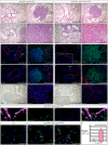
Figure 3. Leaky expression of SPCCreER in alveolar type II cells and not BASCs is associated with alveolar tumors while BADJs retain normal architecture.
(A–F) Histology and immunostaining of lung sections from adult SPCCreER/+; ROSA26mTmG/+ mice without tamoxifen injection. In SPCCreER/+; ROSA26mTmG/+ mice that displayed leaky Cre activity without tamoxifen injection, leaky Cre expression was detected in alveolar type II cells (A) and not BASCs (B). Only alveolar lung tumors were detected in SPCCreER; p53f/f; KrasLSL-G12D/+ mice with leaky Cre activity (D) and BADJs retained normal architecture (C). Tumor cells expressed SPC (type II cell marker) and were highly proliferative (E), suggesting their origin from type II cells. By contrast, no obvious proliferation was observed at BADJs in these mice (F) consistent with their normal structure. Scale bar = 50 µm for panels in each row except (C, D), which is 100 µm.
Figure 4
Figure 4. Induction of alveolar lung tumors in mice with activated EGF signaling in SPC+ cells.
(A–F) Histology of lung sections from SPCrtTA/+; tetO-EGFRL858R adult mice. In the absence of dox chow, lung histology of SPCrtTA/+; tetO-EGFRL858R adult mice was similar to wild-type animals. Once fed with dox chow to activate EGF signaling, SPCrtTA/+; tetO-EGFRL858R mice developed alveolar tumors, but no abnormalities were detected in the airway or BADJ. Interestingly, upon dox withdrawal, tumors regressed and lung histology significantly improved. (G–L) Immunostaining of lung sections from SPCrtTA/+; tetO-EGFRL858R adult mice. Alveolar tumors in SPCrtTA/+; tetO-EGFRL858R adult mice fed with dox expressed SPC (G, J) but not CC10 (G, J). These tumor cells were highly proliferative as assessed by Ki67 staining (L), the entire field of which consists of tumor cells, compared to non-tumor tissues of the lung (K). No obvious hyperplasia was observed in the airway (H) or BADJ (I) in SPCrtTA/+; tetO-EGFRL858R adult mice with active EGFR signaling. Scale bar = 100 µm for each panel.
Figure 5
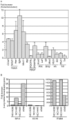
Figure 5. Analysis of major signaling pathways in mouse Kras/p53 tumors and marker expression in human lung cancer cell lines.
(A) Quantitative PCR (qPCR) analysis of RNA extracted from tumors and adjacent non-tumor tissues derived from SPCCreER/+; p53f/f; KrasLSL-G12D/+ adult mice injected with tamoxifen. Target genes of each major signaling pathway were assessed for changes in their expression in tumor versus non-tumor tissues. The numbers represent fold of increase in tumor tissues. Multiple lung tumors from a single mouse and multiple mice were analyzed. Ccnd1 (cyclin D1) was upregulated, consistent with increased Ki67 staining on tumor sections. Notch signaling was consistently upregulated in tumor versus non-tumor tissues whereas Platelet-derived growth factor (PDGF), Wnt, Bone morphogenetic protein (Bmp) and Hedgehog (Hh) pathways showed modest or no apparent changes in target gene expression. Expression levels of Nkx2.1 and Myc were also significantly increased, similar to findings in human adenocarcinoma. Interestingly, expression of Itgb4 (integrin beta 4) was also increased in tumor tissues. Future studies will determine whether Itgb4 plays a role in tumor development. (B) qPCR analysis of RNA extracted from representative human lung cancer cell lines, including NCI-H292 (pulmonary mucoepidermoid carcinoma), NCI-H82 (small cell lung cancer), NCI-H441 (papillary adenocarcinoma of the lung) and NCI-A549 (lung adenocarcinoma). The numbers represent fold of increase in marker expression relative to housekeeping genes. SPC expression was upregulated in the A549 cell line. ITGB4 (integrin beta 4) was also highly expressed in non-small cell lung cancer cell lines compared to small-cell lung cancer cell lines.

References

    1. McErlean A, Ginsberg MS (2011) Epidemiology of lung cancer. Semin Roentgenol 46: 173–177. - PubMed
    1. Ramalingam SS, Owonikoko TK, Khuri FR (2011) Lung cancer: New biological insights and recent therapeutic advances. CA Cancer J Clin 61: 91–112. - PubMed
    1. Murala S, Alli V, Kreisel D, Gelman AE, Krupnick AS (2010) Current status of immunotherapy for the treatment of lung cancer. J Thorac Dis 2: 237–244. - PMC - PubMed
    1. Vallieres E, Peters S, Van Houtte P, Dalal P, Lim E (2011) Therapeutic advances in non-small cell lung cancer. Thorax. - PubMed
    1. Reungwetwattana T, Weroha SJ, Molina JR (2011) Oncogenic Pathways, Molecularly Targeted Therapies, and Highlighted Clinical Trials in Non-Small-Cell Lung Cancer (NSCLC). Clin Lung Cancer. - PubMed