

Jaw expansive lesions: population incidence and CT dentalscan role
- PMID: 23285385
- PMCID: PMC3399186
Jaw expansive lesions: population incidence and CT dentalscan role
Abstract
Aim: The aim of the study is to evaluate the incidence of different expansive lesions and the advantages of the clinical employment of Dentalscan to study bones lesions and to establish a common diagnostic path.
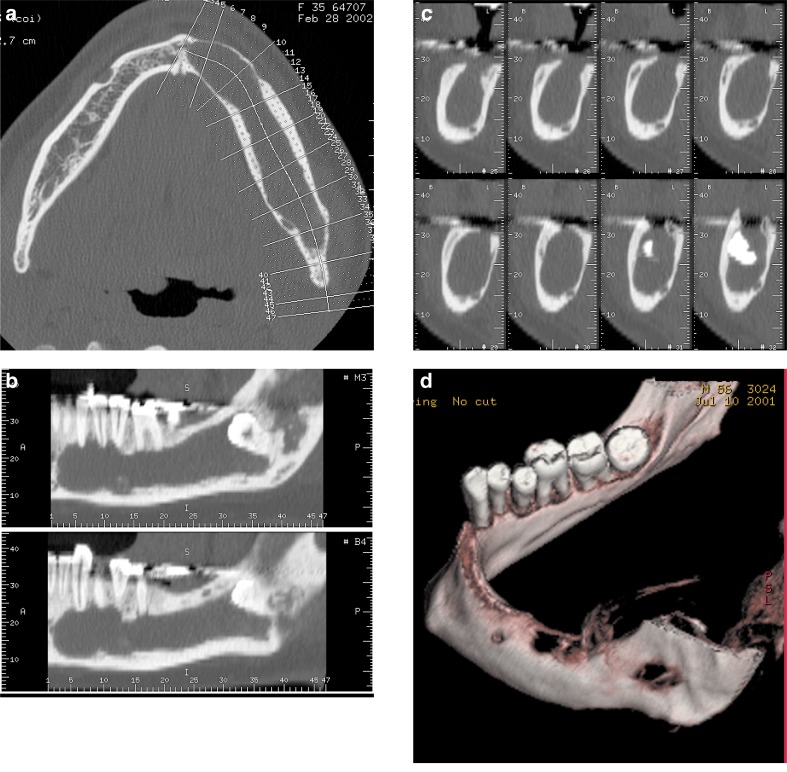
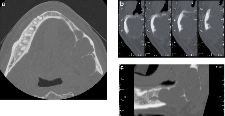
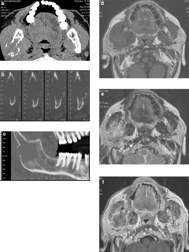
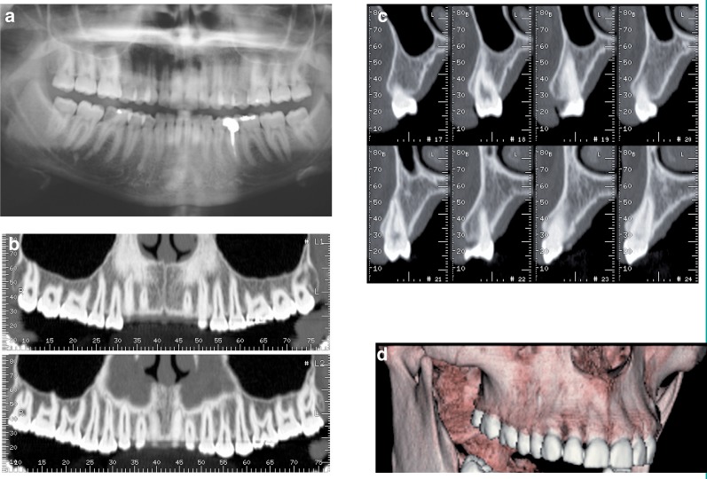
Materials and methods: Since January 2005 to November 2009, 3200 patients, not selected for sex or age, have undergone a CT "Dentalscan" in the department of Diagnostic Imaging, Tor Vergata University Hospital (PTV), a suspect bone pathology was found in 704 of them through the XR-orthopantomograpy (OPT). CT images were obtained with General Electric CT Light Speed multislice. Images were saved in the Advantage Workstation (GE) supported by the "Dentascan" dedicated software and by the 3D software (3D SSD). The protocol was : Slice thickness 1,25 mm, gap 0, matrix 512 × 512, 140 KV and 70 mA. All the lesions were also studied with the dedicated three-dimensional reconstructor 3D SSD. Biopsy for diagnosis was performed on all the lesions, except one (false positive with digital OPT). THE TECHNIQUE SENSITIVITY WAS ASSESSED FOR TWO IMPORTANT CLASSES: benign and malignant lesions.
Results: Through CT Dentascan a detailed evaluation of the jaws lesions and their extension was obtained. 656 patients (93.1 %) out of 704 examined for a suspicious lesion on the orthopantomography had a benign lesion: (127 follicular cysts (18.2 %), 181 radicular cysts (25.1%), 93 non odontogenic cysts (13.2%), 29 fibroma (4.2%), 198 odontomes (28.2%), 24 ameloblastoma (3.6%), 4 brown tumors (0.7%), 47 (6.9%) had malignant lesions: (12 carcinoma (1.7%), 29 metastasis (4.3%), 6 sarcoma (0.8%), 1 Dentascan CT resulted to be negative (1 false positive of digital OPT). The sensitivity of the technique for both groups was 99% for benign lesions and 98% for malign lesions.
Conclusions: CT Dentascan characteristics suggest to consider these techniques as the gold standard for the evaluation of jaw expansive lesions and the support of surgical planning.
Scopo del lavoro: Scopo del lavoro è valutare l’incidenza delle varie lesioni espansive e i possibili vantaggi nell’impiego clinico del Dentascan nella valutazione delle lesioni ossee anche nell’ottica di una standardizzazione dell’iter diagnostico.
Materiali e metodi: Nel periodo compreso tra gennaio 2005 e novembre 2009, presso il Dipartimento di Diagnostica per immagini del Policlinico Universitario di Tor Vergata (PTV), sono stati sottoposti a studio con TC “Dentascan” 3200 pazienti, non selezionati per sesso e per età, di cui 704 con patologia ossea sospetta all’indagine radiografica tradizionale eseguita con ortopantomografia (OPT). Gli esami TC sono stati ottenuti con apparecchio General Electric CT Light Speed multislice e sono stati successivamente trasferiti alla stazione di lavoro “Advantage windows” (GE) supportata dal software di ricostruzione dedicato “Dentascan” oltre che dal software di ricostruzione tridimensionale (3D SSD). È stato impiegato un protocollo con spessore di strato di 1,25 mm, gap 0, matrice 512 × 512, 140 KV e 70 mA.
Tutte le lesioni ossee sono state studiate anche con ricostruzione tridimensionale 3D SSD. In tutti i casi tranne uno (falso positivo all’OPT digitale) si è reso necessario l’esame bioptico per una diagnosi di certezza.
È stata inoltre valutata la sensibilità della tecnica per due grandi gruppi: lesione benigna e lesione maligna.
Risultati: La TC Dentascan ha consentito in tutti i casi uno studio dettagliato delle lesioni riscontrate a livello dell’osso mascellare e della mandibola e la loro estensione. Di 704 (22%) pazienti esaminati con lesione ossea sospetta all’ortopantomografia, 656 (93.1%) avevano alterazioni di tipo benigno, di cui 127 cisti follicolari (18.2%), 181 cisti radicolari (25.1%), 93 cisti non odontogene (13.2%), 29 fibromi (4.2%), 198 odontomi (28.2%), 24 ameloblastomi (3.6%), 4 tumori bruni (0.7%), 47 (6.9%) alterazioni maligne: (12 carcinomi (1.7%), 29 metastasi (4.3%), 6 sarcomi (0.8%), in 1 paziente la TC Dentascan è risultata negativa (1 falso positivo all’OPT digitale).
La sensibilità della tecnica per due grandi gruppi è stata del 99% per le lesioni benigne e del 98% per le lesioni maligne.
Conclusione: Le caratteristiche intrinseche della tecnica la propongono come tecnica di scelta nella valutazione dei processi espansivi dei mascellari facendo da supporto anche alla pianificaizone chirurgica.
Keywords: dentalscan; expansive lesions.
Figures
References
-
- Abrahams JJ. Dental CT Imaging: a look at the jaw. Radiology. 2001;219:334–345. - PubMed
-
- Abrahams JJ, Berger S. Inflammatory disease of the jaw: appearance on reformatted CT images. AJR Am J Roentgenol. 1998;170:1085–1091. - PubMed
-
- Abrahams JJ, Frisoli JK, Dembner J. Anatomy of the jaw, dentition, and related region. Semin Ultrasound CT MR. 1995;16:453–467. - PubMed
-
- Abrahams JJ. The role of diagnostic imaging in dental implantology. Radiol Clin North Am. 1993;31:163–180. - PubMed
-
- Au-yeung KM, Ahuja AT, Ching ASC. Metreweli C.: DentaScan in Oral Imaging. Clinical Radiology. 2001;56:700–713. - PubMed
LinkOut - more resources
Full Text Sources
Miscellaneous