

Registration and analysis of white matter group differences with a multi-fiber model
- PMID: 23286145
- PMCID: PMC3671390
- DOI: 10.1007/978-3-642-33454-2_39
Registration and analysis of white matter group differences with a multi-fiber model
Abstract
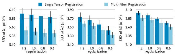
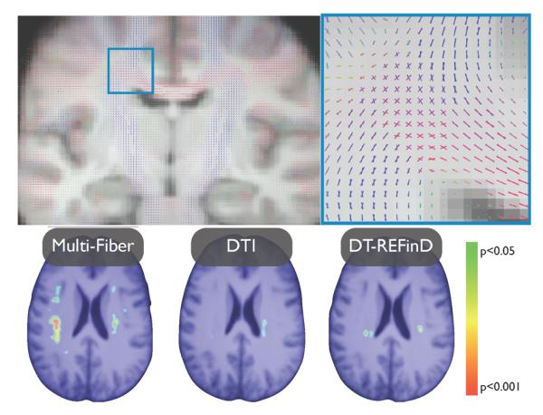
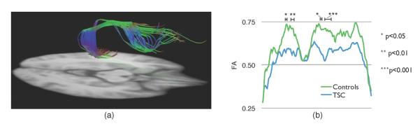
Diffusion magnetic resonance imaging has been used extensively to probe the white matter in vivo. Typically, the raw diffusion images are used to reconstruct a diffusion tensor image (DTI). The incapacity of DTI to represent crossing fibers leaded to the development of more sophisticated diffusion models. Among them, multi-fiber models represent each fiber bundle independently, allowing the direct extraction of diffusion features for population analysis. However, no method exists to properly register multi-fiber models, seriously limiting their use in group comparisons. This paper presents a registration and atlas construction method for multi-fiber models. The validity of the registration is demonstrated on a dataset of 45 subjects, including both healthy and unhealthy subjects. Morphometry analysis and tract-based statistics are then carried out, proving that multi-fiber models registration is better at detecting white matter local differences than single tensor registration.
Figures
References
-
- Assaf Y, Basser P. Composite hindered and restricted model of diffusion (charmed) mr imaging of the human brain. Neuroimage. 2005;27(1):48–58. - PubMed
-
- Banerjee A, Merugu S, Dhillon I, Ghosh J. Clustering with bregman divergences. The Journal of Machine Learning Research. 2005;6:1705–1749.
-
- Barmpoutis A, Vemuri B. Groupwise registration and atlas construction of 4th-order tensor fields using the r+ riemannian metric. Medical Image Computing and Computer-Assisted Intervention–MICCAI 2009. 2009:640–647. - PubMed
-
- Bergmann O, Kindlmann G, Peled S, Westin C. Two-tensor fiber tractography. Biomedical Imaging: From Nano to Macro, 2007. ISBI 2007. 4th IEEE International Symposium on; IEEE; 2007. pp. 796–799.
Publication types
MeSH terms
Grants and funding
LinkOut - more resources
Full Text Sources
Medical