. 2013 Jan 22;52(3):488-96.
doi: 10.1021/bi301294d. Epub 2013 Jan 9.

Characterizing the importance of the biotin carboxylase domain dimer for Staphylococcus aureus pyruvate carboxylase catalysis

Affiliations

Characterizing the importance of the biotin carboxylase domain dimer for Staphylococcus aureus pyruvate carboxylase catalysis

Linda P C Yu et al. Biochemistry. .

Abstract

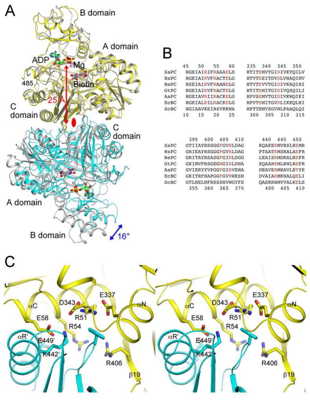
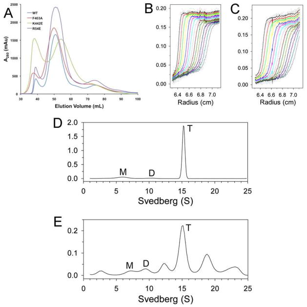
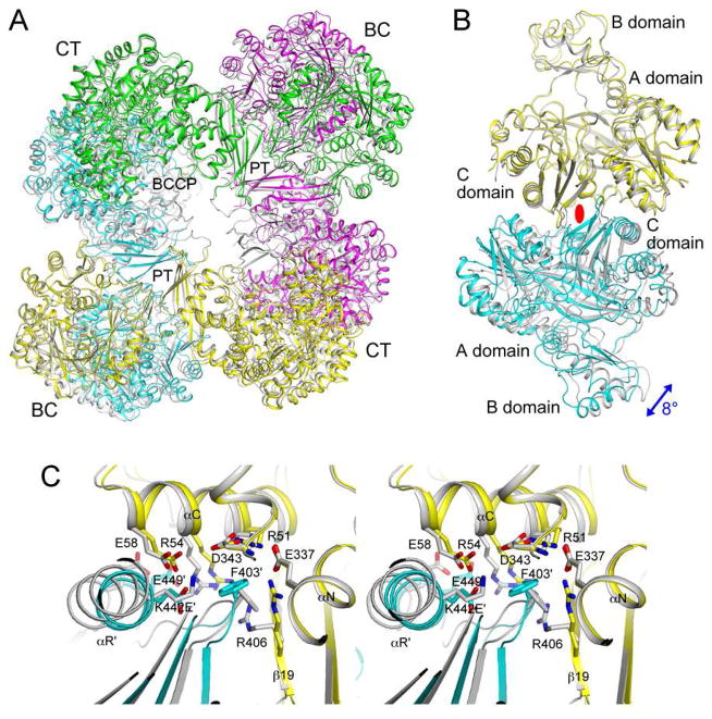
Biotin carboxylase (BC) is a conserved component among biotin-dependent carboxylases and catalyzes the MgATP-dependent carboxylation of biotin, using bicarbonate as the CO₂ donor. Studies with Escherichia coli BC have suggested long-range communication between the two active sites of a dimer, although its mechanism is not well understood. In addition, mutations in the dimer interface can produce stable monomers that are still catalytically active. A homologous dimer for the BC domain is observed in the structure of the tetrameric pyruvate carboxylase (PC) holoenzyme. We have introduced site-specific mutations into the BC domain dimer interface of Staphylococcus aureus PC (SaPC), equivalent to those used for E. coli BC, and also made chimeras replacing the SaPC BC domain with the E. coli BC subunit (EcBC chimera) or the yeast ACC BC domain (ScBC chimera). We assessed the catalytic activities of these mutants and characterized their oligomerization states by gel filtration and analytical ultracentrifugation experiments. The K442E mutant and the ScBC chimera disrupted the BC dimer and were catalytically inactive, while the F403A mutant and the EcBC chimera were still tetrameric and retained catalytic activity. The R54E mutant was also tetrameric but was catalytically inactive. Crystal structures of the R54E, F403A, and K442E mutants showed that they were tetrameric in the crystal, with conformational changes near the mutation site as well as in the tetramer organization. We have also produced the isolated BC domain of SaPC. In contrast to E. coli BC, the SaPC BC domain is monomeric in solution and catalytically inactive.


Figures

Figure 1
Figure 1. Conserved BC domain dimer interface for SaPC and E. coli BC
(A). Overlay of the structures of the BC domain dimer of SaPC free enzyme (in yellow and cyan for the two monomers) (25) and the E. coli BC dimer (in gray) in complex with ADP (in green), Mg2+ (pink sphere), bicarbonate (in black), and biotin (in pink) (14). The blue arrow indicates the difference in orientation for the second monomer of the dimer between the two structures, and the red arrow indicates the distance from the dimer interface to the active site. The two-fold axis of the dimer is indicated with the red oval. (B). Alignment of amino acid sequences in the dimer interface region for SaPC, human PC (HsPC), RePC, Geobacillus thermodenitrificans PC (GtPC), Aquifex aeolicus PC (AaPC), E. coli BC (EcBC) and the BC domain of yeast ACC (ScBC). Residues having important interactions in the interface are shown in red. Residue numbers at the top are for SaPC (in human PC numbering), and those at the bottom are for E. coli BC. (C). Detailed interactions at the BC domain dimer interface of SaPC. Residues Arg54, Glu58, Phe403 and Lys442 were selected for mutagenesis. The primed residue numbers indicate the second monomer. All structure figures were produced with PyMOL (www.pymol.org).
Figure 2
Figure 2. Oligomerization states of the dimer interface mutants in solution
(A). Gel filtration profiles (from an S300 column) for wild-type SaPC and the dimer interface mutants. (B). Sedimentation velocity AUC data for wild-type SaPC at 1.5 μM concentration. The observed data are shown as open circles, and the theoretical fit to the data based on a rapid monomer-dimer-tetramer association model is shown as the curves. (C). Sedimentation velocity AUC data for the K442E mutant at 1.5 μM concentration. (D). Size distributions of wild-type SaPC in solution at 1 mg/ml concentration, based on the best-fit results by the continuous size distribution analysis (30). M: monomer, D: dimer, T: tetramer. (E). Size distributions of the EcBC chimera in solution at 1 mg/ml concentration.
Figure 3
Figure 3
The R54E mutant has only small structural differences compared to wild-type SaPC. (A). Overlay of the structures of the R54E mutant tetramer (in color for the four monomers, yellow, magenta, cyan and green) and wild-type SaPC (gray). The superposition is based on the BC domain of the first monomer (yellow). (B). Overlay of the structures of the R54E mutant BC domain dimer (yellow and cyan) and wild-type SaPC dimer (gray). The blue arrow indicates the difference in orientation for the second monomer of the dimer between the two structures. The two-fold axis of the dimer is indicated with the red oval. (C). Close-up of the BC domain dimer interface near the mutation site.
Figure 4
Figure 4
The F403A mutant shows large structural differences compared to wild-type SaPC. (A). Overlay of the structures of the F403A mutant tetramer (in color) and wild-type SaPC (gray). The superposition is based on the BC domain of the first monomer (yellow). (B). Overlay of the structures of the F403A mutant BC domain dimer (yellow and cyan) and wild-type SaPC dimer (gray). (C). Close-up of the BC domain dimer interface near the mutation site.
Figure 5
Figure 5
The K442E mutant is a tetramer in the crystal but shows large structural differences compared to wild-type SaPC. (A). Overlay of the structures of the K442E mutant tetramer (in color) and wild-type SaPC (gray). The superposition is based on the BC domain of the first monomer (yellow). (B). Overlay of the structures of the K442E mutant BC domain dimer (yellow and cyan) and wild-type SaPC dimer (gray). (C). Close-up of the BC domain dimer interface near the mutation site.

References

    1. Wakil SJ, Stoops JK, Joshi VC. Fatty acid synthesis and its regulation. Ann Rev Biochem. 1983;52:537–579. - PubMed
    1. Cronan JE, Jr, Waldrop GL. Multi-subunit acetyl-CoA carboxylases. Prog Lipid Res. 2002;41:407–435. - PubMed
    1. Tong L. Acetyl-coenzyme A carboxylase: crucial metabolic enzyme and attractive target for drug discovery. Cell Mol Life Sci. 2005;62:1784–1803. - PMC - PubMed
    1. Jitrapakdee S, St Maurice M, Rayment I, Cleland WW, Wallace JC, Attwood PV. Structure, mechanism and regulation of pyruvate carboxylase. Biochem J. 2008;413:369–387. - PMC - PubMed
    1. Tong L. Structure and function of biotin-dependent carboxylases. Cell Mol Life Sci 2012 - PMC - PubMed

Publication types

MeSH terms

LinkOut - more resources