Review
. 2013 Mar 15;8(3):473-87.
doi: 10.1021/cb3005325. Epub 2013 Jan 8.

Revealing nature's synthetic potential through the study of ribosomal natural product biosynthesis

Affiliations
Review

Revealing nature's synthetic potential through the study of ribosomal natural product biosynthesis

Kyle L Dunbar et al. ACS Chem Biol. .

Erratum in

  • ACS Chem Biol. 2013 May 17;8(5):1083

Abstract

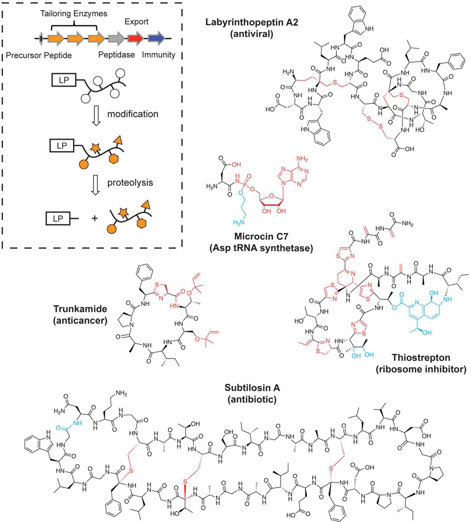
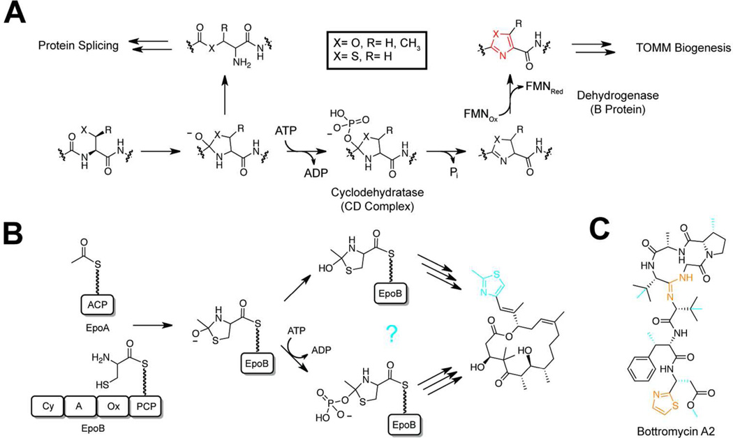
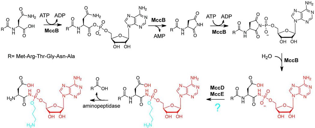
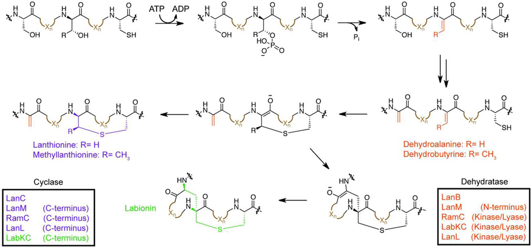
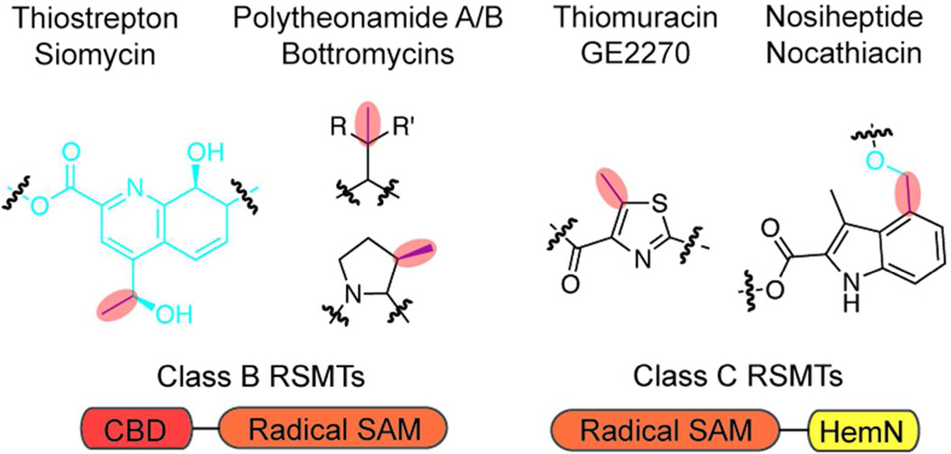
Ribosomally synthesized posttranslationally modified peptides (RiPPs) are a rapidly growing class of natural products with diverse structures and activities. In recent years, a great deal of progress has been made in elucidating the biosynthesis of various RiPP family members. As with the study of nonribosomal peptide and polyketide biosynthetic enzymes, these investigations have led to the discovery of entirely new biological chemistry. With each unique enzyme investigated, a more complex picture of Nature's synthetic potential is revealed. This Review focuses on recent reports (since 2008) that have changed the way that we think about ribosomal natural product biosynthesis and the enzymology of complex bond-forming reactions.


Figures

Figure 1
Figure 1
Example of RiPP natural product diversity. The general strategy for RiPP biosynthesis is illustrated in the generic gene cluster and schematic below. The functional assignment for each of the open reading frames is displayed. An array of ribosomal natural products generated from this strategy is displayed along with their molecular target. In all figures within this review, the red moieties represent posttranslational modifications where the tailoring enzyme(s) responsible have been characterized. The modification mechanisms for the blue moieties are unknown and are shown to highlight additional chemistry that remains unexplored in RiPP modification enzymes.
Figure 2
Figure 2
Cyclizations via backbone activation. a) A mechanistic proposal for the biosynthesis of azoline heterocycles in TOMM natural products. The first step is shared with the mechanism of protein splicing. Following phosphorylation and elimination of phosphate, a FMN-dependent dehydrogenation affords the aromatic azole (red). b) Two possible strategies are presented for the biosynthesis of azoline heterocycles in PKS/NRPS natural products (e.g. epothilone). The top route is ATP-independent while the bottom follows the mechanism of the TOMM cyclodehydratase complex. As the mechanism for the Cy-dependent azoline formation is unknown the moiety is shown in blue. c) The structure of bottromycin A2 is shown with the putative YcaO-installed modifications highlighted in orange. Modifications installed by uncharacterized mechanisms are shown in blue.
Figure 3
Figure 3
Microcin C7 biosynthesis. The mechanism of the MccB-catalyzed phosphoramidate bond installation is shown with the final posttranslational modification of the precursor peptide displayed in red. Addition of the aminopropyl group (blue) to the phosphoramidate moiety is proposed to be catalyzed by MccD and/or MccE enzymes. Upon entering a target cell, the N-terminal six amino acids are proteolyically removed to afford the bioactive compound.
Figure 4
Figure 4
Lanthionine ring formation in lantipeptides. The mechanism of dehydration of Ser/Thr residues to generate dehydroalanine/dehydrobutyrine moieties (orange) in lantipeptides is displayed. In all studied cases, dehydration is ATP-dependent and proceeds through a phosphorylated intermediate. Subsequently, a cyclase domain catalyzes a Michael-type addition to form the lanthionine rings (purple). In a subclass of lantipeptides, a second Michael-addition occurs to generate a labionin ring (green). In all cases, the enzymes responsible for each transformation are displayed in the same color as the modification they install.
Figure 5
Figure 5
Strategies for natural product macrocyclization. a) Cyanobactin biosynthetic scheme. Oxazoline (OxH; purple) and thiazole (Thz; purple) heterocycles are installed on cyanobactin precursor peptides by PatD (cyclodehydratase) and the N-terminal oxidation domain of PatG. After heterocycle installation, the peptide is proteolyzed by PatA and PatG, the latter also catalyzing macrocyclization (red). b) The PatG macrocyclization mechanism is displayed with the catalytic triad shown. The residues important for recognition in the C-terminal AYDG recognition sequence are colored orange. c) As a comparison, a mechanism for the DEBS thioesterase domain (non-RiPP) is displayed. This mechanism is conserved in a majority of the characterized PKS and NRPS thioesterases.
Figure 6
Figure 6
[4+2] cycloadditions in natural product biosynthesis. a) Two potential mechanisms for installing the pyridine moiety of thiocillin I (red) are displayed. Starting from the tautomer form, the orange arrows demonstrate a concerted Diels-Alder mechanism, while the purple arrows follow a stepwise, polar mechanism. The shared step of each mechanism is displayed as a dashed black arrow. After the [4+2] cycloaddition is complete, the elimination of water and the leader peptide (LP) affords the central pyridine ring. The colored circles on the tautomer form of the non-cyclized intermediate indicate positions where dual 13C-labeling could distinguish between stepwise and concerted cyclization mechanisms. b) The SpnF-catalyzed cycloaddition of spinosin A (red) is displayed. The colored circles indicate positions where dual 2H-labeling could be used for establishing the precise mechanism.
Figure 7
Figure 7
The mechanism of MIA formation in nosiheptide biosynthesis. a) The structure of nosiheptide is displayed with the indolic acid ring highlighted in red. The uncharacterized radical SAM methyltransferase, NosA, and an as of yet unidentified protein, methylate and oxidize MIA to afford the second attachment point (blue moiety). b) The proposed mechanism of indolic acid formation by the radical SAM protein NosL is shown. The structures of the intermediates/products enclosed in the dotted circles were detected in reaction mixtures. The colored asterisks denote 13C-labeled positions that were used to demonstrate the fate of the carbon backbone during the rearrangement.
Figure 8
Figure 8
Subtilosin biosynthesis. a) The general biosynthetic scheme for subtilosin is shown. Residues involved in thioether crosslinking are colored (red, donor residue; purple, receiver residue). The protein(s) responsible for leader peptide (LP) cleavage and macrocyclization (blue) have not been characterized. b) A proposed mechanism of thioether formation is presented. The first Fe-S cluster is involved in radical generation while the second cluster serves as a one-electron oxidant of the cysteine thiol.
Figure 9
Figure 9
Radical SAM-dependent methylations in RiPPs. Shown are several examples of RSMTdependent RiPP modifications (red highlight). Shown below each structure is the class of RSMT and its domain architecture (CBD, cobalamin-binding domain). The blue moieties are installed by uncharacterized enzymes. R= H, R’= Ph or R= Me, R’= Me/Et.
Figure 10
Figure 10
Cyanobactin prenylation. a) The two forms of O-prenylation are displayed along with examples of natural products containing each modification. b) The LynF prenylation mechanism. After reverse O-prenylation of tyrosine, the intermediate undergoes a Claisen rearrangement to afford the C-prenylated product. As the product of the Lyn cluster has not been identified, the predicted product is displayed without stereochemistry. The DMAPP derived prenyl groups are shown in red.
Figure 11
Figure 11
The structure of sublancin. The modification installed by SunS (S-glycosyltransferase) is shown in red.
Figure 12
Figure 12
C-terminal amidation of thiopeptides. The putative mechanism for C-terminal amide formation is displayed for nosiheptide a) and thiostrepton b). The proposed transformation catalyzed by TsrC is boxed. Tables listing the other thiopeptides that utilize each mechanism, along with the pertinent protein, are listed on the right.

References

    1. Harvey AL. Natural products in drug discovery. Drug Discov. Today. 2008;13:894–901. - PubMed
    1. Newman DJ, Cragg GM. Natural products as sources of new drugs over the 30 years from 1981 to 2010. J. Nat. Prod. 2012;75:311–335. - PMC - PubMed
    1. Wilson RM, Danishefsky SJ. Small molecule natural products in the discovery of therapeutic agents: the synthesis connection. J. Org. Chem. 2006;71:8329–8351. - PubMed
    1. Razzak M, De Brabander JK. Lessons and revelations from biomimetic syntheses. Nat. Chem. Biol. 2011;7:865–875. - PubMed
    1. Falconer SB, Czarny TL, Brown ED. Antibiotics as probes of biological complexity. Nat. Chem. Biol. 2011;7:415–423. - PubMed

Publication types