. 2013 Jan 3;20(1):1.
doi: 10.1186/1423-0127-20-1.

Concurrent exposure to a dectin-1 agonist suppresses the Th2 response to epicutaneously introduced antigen in mice

Affiliations

Concurrent exposure to a dectin-1 agonist suppresses the Th2 response to epicutaneously introduced antigen in mice

Jing-Yi Lin et al. J Biomed Sci. .

Abstract

Background: Epicutaneous sensitization with protein allergen that induces predominant Th2 responses is an important sensitization route in atopic dermatitis. Fungal components have been shown to modulate Th cell differentiation. However, the effects of fungal components on epicutaneous sensitization are unclear.

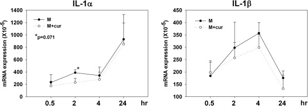
Results: In this study, we showed that co-administration of curdlan, a dectin-1 agonist, during epicutaneous ovalbumin sensitization of BALB/c mice decreased the IL-5 and IL-13 levels in supernatants of lymph node cell ovalbumin reactivation cultures. Mechanistically, curdlan co-administration decreased IL-4 and IL-1β expressions in draining lymph nodes. Curdlan co-administration also lower the migration of langerin+ CD103- epidermal Langerhans cells into draining lymph nodes at 96 hours post-sensitization which might be attributed to decreased expressions of IL-18 and IL-1β in patched skin. Moreover, adoptive transfer of CFSE-labeled transgenic CD4 T cells confirmed that curdlan co-administration decreased the proliferation and IL-4-production of ovalbumin -specific T cells primed by epidermal Langerhans cells.

Conclusions: These results indicated that concurrent exposure to a dectin-1 agonist suppresses the epicutaneously induced Th2 response by modulating the cytokine expression profiles in draining LNs and the migration of epidermal Langerhans cells. These results highlight the effects of fungal components on epicutaneous allergen sensitization in atopic diseases.


Figures

Figure 1
Figure 1
Co-administration of dectin-1 agonists suppresses the epicutaneously OVA-induced Th2 immune response. (A) Groups of BALB/c mice (n=5) were sensitized by patch application with OVA plus doses of curdlan or OVA alone on day1-5. (B) Groups of BALB/c mice (n=5) were sensitized by patch application with OVA plus zymosan, OVA plus Pam3CSK4 and doses of curdlan, or OVA alone on day 1–5. Ten days after the start of the sensitization course, draining LNs were obtained. The IL-5 and IL-13 contents in supernatants of in vitro OVA reactivation culture of LN cells were determined by ELISA. Net concentrations (concentration in the absence of OVA subtracted from concentration in the presence of OVA) were presented. Data are representative of at least three independent experiments.
Figure 2
Figure 2
Co-administration of curdlan with OVA influences the cytokine expression profiles in draining LNs. Groups of BALB/c mice were sensitized by patch application with OVA plus curdlan or OVA alone. Draining LNs were obtained at 24, 48, 72 and 96 hours after sensitization. Total RNA extraction, cDNA preparation and quantitative real-time PCR for various cytokines were performed. The relative cytokine mRNA expression of each group was normalized to its β-actin expression. Results are shown as means and standard deviations of pooled data from five independent experiments. P values was calculated by Wilcoxon Rank Sum test.
Figure 3
Figure 3
Co-administration of curdlan decreases the number of epidermal LC in draining LNs at 96 hours post-epicutaneous sensitization. (A) Groups of BALB/c mice were sensitized by patch application with OVA plus curdlan or OVA alone. 24 and 96 hours after patch application, draining LNs were obtained and prepared for positive selection for DCs. Intracellular staining for langerin and surface staining for CD103 were performed. Langerin+ CD103- cells represent epidermal LCs. The results were shown as mean ± SD of pooled data from four independent experiments. (B) 96 hours after patch application, intracellular staining for langerin and surface staining for CD103 and various costimulatory molecules were performed. Langerin+ CD103- cells were gated. Expression levels of CD86 and CD40 were shown. OVA group (gray line), OVA plus curdlan group (black line), isotype control (gray dotted line).
Figure 4
Figure 4
Co-administration of curdlan with OVA influences the cytokine expression profiles in patched skin. Groups of BALB/c mice were sensitized by patch application with OVA plus curdlan or OVA alone. Patched skins were excised 2, 4 and 24 hours after patch application. Total RNA extract, cDNA preparation and quantitative real-time PCR for various cytokines were performed. The relative cytokine mRNA expression of each group was normalized to its β-actin expression. Results are shown as means and standard deviations of pooled data from four independent experiments.
Figure 5
Figure 5
Presence of curdlan inin vitroculture decreases the IL-1α and IL-1β expressions of primary keratinocytes. Skin was obtained from naïve BALB/c mice and prepared into epidermal cell suspension. Keratinocytes were purified by negative selection with MHC class II bead. In vitro culture of primary keratinocytes in the absence or presence of curdlan (50 μg/ml) were performed. Keratinocytes were harvested 0.5, 2, 4 and 24 hours after the start of culture. Measurement of various cytokines were performed similar to Figure 4.
Figure 6
Figure 6
Co-administration of curdlan decreases the proliferation and IL-4 production of specific T cells primed by epidermal LCs. CFSE-labeled CD4 T cell from DO.11.10 mice were intravenously transferred into groups of BALB/c mice three days after they received patch application with OVA plus curdlan or OVA alone. Draining LNs were obtained three days after transfer. (A) After staining with CD4, flow cytometric analysis was performed. CD4+ CFSE+ cells were gated. (B) Pooled results of percentages of divided, labeled CD4 T cells of individual mouse from three independent experiments (n=12) were shown. (C) With intracellular staining for IL-4, pooled results of numbers of IL-4-producing, labeled CD4 T cells of individual mouse from two independent experiments were shown (n=8).

Similar articles

Cited by

References

    1. Ngoc PL, Gold DR, Tzianabos AO, Weiss STCeledon JC. Cytokines, allergy, and asthma. Curr Opin Allergy Clin Immunol. 2005;5:161–166. doi: 10.1097/01.all.0000162309.97480.45. - DOI - PubMed
    1. Spergel JMPaller AS. Atopic dermatitis and the atopic march. J Allergy Clin Immunol. 2003;112:S118–S127. doi: 10.1016/j.jaci.2003.09.033. - DOI - PubMed
    1. Santamaria Babi LF, Picker LJ, Perez Soler MT, Drzimalla K, Flohr P, Blaser KHauser C. Circulating allergen-reactive T cells from patients with atopic dermatitis and allergic contact dermatitis express the skin-selective homing receptor, the cutaneous lymphocyte-associated antigen. J Exp Med. 1995;181:1935–1940. doi: 10.1084/jem.181.5.1935. - DOI - PMC - PubMed
    1. Teraki Y, Hotta TShiohara T. Increased circulating skin-homing cutaneous lymphocyte-associated antigen (CLA)+ type 2 cytokine-producing cells, and decreased CLA+ type 1 cytokine-producing cells in atopic dermatitis. Br J Dermatol. 2000;143:373–378. doi: 10.1046/j.1365-2133.2000.03665.x. - DOI - PubMed
    1. Wang LF, Lin JY, Hsieh KHLin RH. Epicutaneous exposure of protein antigen induces a predominant Th2-like response with high IgE production in mice. J Immunol. 1996;156:4077–4082. - PubMed

Publication types

MeSH terms