

Partial functional conservation of IRX10 homologs in physcomitrella patens and Arabidopsis thaliana indicates an evolutionary step contributing to vascular formation in land plants
- PMID: 23286876
- PMCID: PMC3543728
- DOI: 10.1186/1471-2229-13-3
Partial functional conservation of IRX10 homologs in physcomitrella patens and Arabidopsis thaliana indicates an evolutionary step contributing to vascular formation in land plants
Abstract
Background: Plant cell walls are complex multicomponent structures that have evolved to fulfil an essential function in providing strength and protection to cells. Hemicelluloses constitute a key component of the cell wall and recently a number of the genes thought to encode the enzymes required for its synthesis have been identified in Arabidopsis. The acquisition of hemicellulose synthesis capability is hypothesised to have been an important step in the evolution of higher plants.
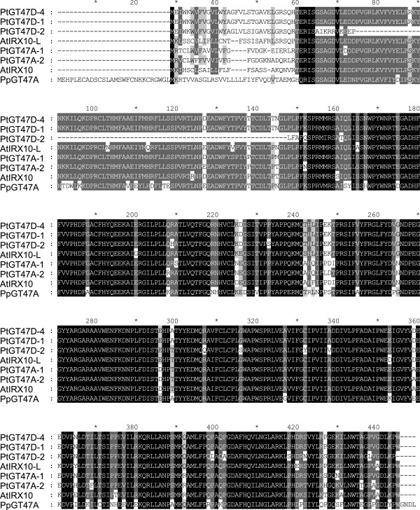
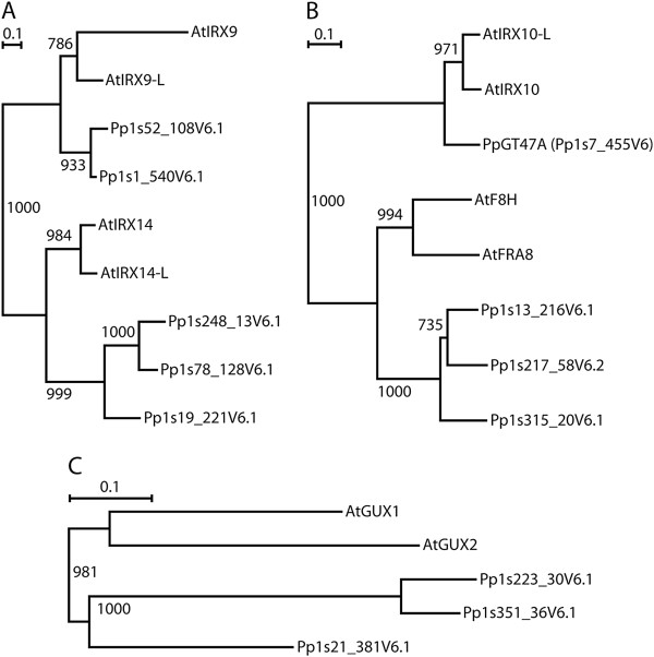
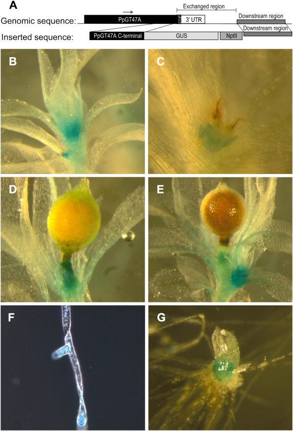
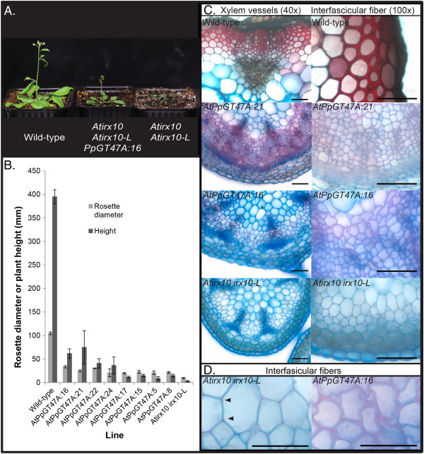
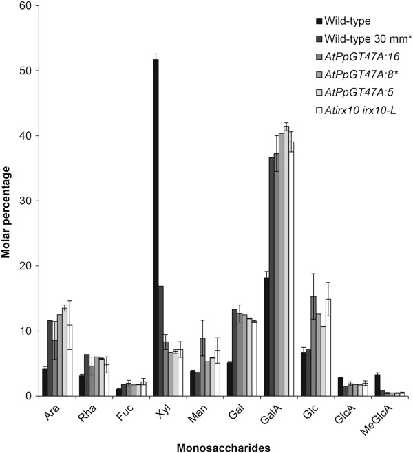
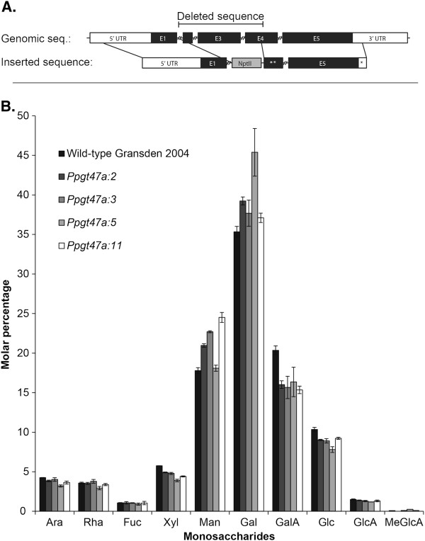
Results: Analysis of the Physcomitrella patens genome has revealed the presence of homologs for all of the Arabidopsis glycosyltransferases including IRX9, IRX10 and IRX14 required for the synthesis of the glucuronoxylan backbone. The Physcomitrella IRX10 homolog is expressed in a variety of moss tissues which were newly formed or undergoing expansion. There is a high degree of sequence conservation between the Physcomitrella IRX10 and Arabidopsis IRX10 and IRX10-L. Despite this sequence similarity, the Physcomitrella IRX10 gene is only able to partially rescue the Arabidopsis irx10 irx10-L double mutant indicating that there has been a neo- or sub-functionalisation during the evolution of higher plants. Analysis of the monosaccharide composition of stems from the partially rescued Arabidopsis plants does not show any significant change in xylose content compared to the irx10 irx10-L double mutant. Likewise, knockout mutants of the Physcomitrella IRX10 gene do not result in any visible phenotype and there is no significant change in monosaccharide composition of the cell walls.
Conclusions: The fact that the Physcomitrella IRX10 (PpGT47A) protein can partially complement an Arabidopsis irx10 irx10-L double mutant suggests that it shares some function with the Arabidopsis proteins, but the lack of a phenotype in knockout lines shows that the function is not required for growth or development under normal conditions in Physcomitrella. In contrast, the Arabidopsis irx10 and irx10 irx10-L mutants have strong phenotypes indicating an important function in growth and development. We conclude that the evolution of vascular plants has been associated with a significant change or adaptation in the function of the IRX10 gene family.
Figures
Similar articles
-
Arabidopsis thaliana IRX10 and two related proteins from psyllium and Physcomitrella patens are xylan xylosyltransferases.Plant J. 2014 Oct;80(2):207-15. doi: 10.1111/tpj.12641. Epub 2014 Sep 15. Plant J. 2014. PMID: 25139408
-
Analysis of the Arabidopsis IRX9/IRX9-L and IRX14/IRX14-L pairs of glycosyltransferase genes reveals critical contributions to biosynthesis of the hemicellulose glucuronoxylan.Plant Physiol. 2010 Jun;153(2):542-54. doi: 10.1104/pp.110.154971. Epub 2010 Apr 27. Plant Physiol. 2010. PMID: 20424005 Free PMC article.
-
Site-directed mutagenesis of IRX9, IRX9L and IRX14 proteins involved in xylan biosynthesis: glycosyltransferase activity is not required for IRX9 function in Arabidopsis.PLoS One. 2014 Aug 13;9(8):e105014. doi: 10.1371/journal.pone.0105014. eCollection 2014. PLoS One. 2014. PMID: 25118690 Free PMC article.
-
Cell wall biology of the moss Physcomitrium patens.J Exp Bot. 2022 Jul 16;73(13):4440-4453. doi: 10.1093/jxb/erac122. J Exp Bot. 2022. PMID: 35348679 Review.
-
Plant Cell Wall Proteomes: The Core of Conserved Protein Families and the Case of Non-Canonical Proteins.Int J Mol Sci. 2022 Apr 12;23(8):4273. doi: 10.3390/ijms23084273. Int J Mol Sci. 2022. PMID: 35457091 Free PMC article. Review.
Cited by
-
β-1,4-Xylan backbone synthesis in higher plants: How complex can it be?Front Plant Sci. 2023 Jan 11;13:1076298. doi: 10.3389/fpls.2022.1076298. eCollection 2022. Front Plant Sci. 2023. PMID: 36714768 Free PMC article.
-
Hemicellulose biosynthesis.Planta. 2013 Oct;238(4):627-42. doi: 10.1007/s00425-013-1921-1. Epub 2013 Jun 26. Planta. 2013. PMID: 23801299 Review.
-
Ceratopteris richardii (C-fern): a model for investigating adaptive modification of vascular plant cell walls.Front Plant Sci. 2013 Sep 23;4:367. doi: 10.3389/fpls.2013.00367. eCollection 2013. Front Plant Sci. 2013. PMID: 24065974 Free PMC article.
-
Immuno and Affinity Cytochemical Analysis of Cell Wall Composition in the Moss Physcomitrella patens.Front Plant Sci. 2016 Mar 8;7:248. doi: 10.3389/fpls.2016.00248. eCollection 2016. Front Plant Sci. 2016. PMID: 27014284 Free PMC article.
-
Arabidopsis VASCULAR-RELATED UNKNOWN PROTEIN1 regulates xylem development and growth by a conserved mechanism that modulates hormone signaling.Plant Physiol. 2014 Apr;164(4):1991-2010. doi: 10.1104/pp.114.236406. Epub 2014 Feb 24. Plant Physiol. 2014. PMID: 24567189 Free PMC article.
References
-
- Brown RM. Cellulose microfibril assembly and orientation - recent developments. J Cell Sci. 1985;2:13–32. - PubMed
-
- Ligrone R, Vaughn KC, Renzaglia KS, Knox JP, Duckett JG. Diversity in the distribution of polysaccharide and glycoprotein epitopes in the cell walls of bryophytes: new evidence for the multiple evolution of water-conducting cells. New Phytol. 2002;156:491–508. doi: 10.1046/j.1469-8137.2002.00538.x. - DOI - PubMed
Publication types
MeSH terms
Substances
LinkOut - more resources
Full Text Sources
Other Literature Sources
Molecular Biology Databases
