

Molecular changes during egg activation
- PMID: 23287037
- PMCID: PMC3931425
- DOI: 10.1016/B978-0-12-416024-8.00010-6
Molecular changes during egg activation
Abstract
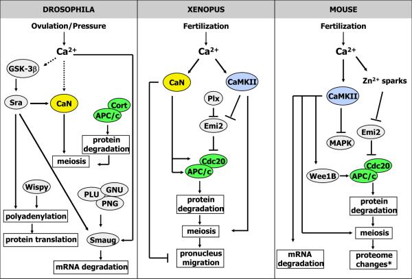
Egg activation is the final transition that an oocyte goes through to become a developmentally competent egg. This transition is usually triggered by a calcium-based signal that is often, but not always, initiated by fertilization. Activation encompasses a number of changes within the egg. These include changes to the egg's membranes and outer coverings to prevent polyspermy and to support the developing embryo, as well as resumption and completion of the meiotic cell cycle, mRNA polyadenylation, translation of new proteins, and the degradation of specific maternal mRNAs and proteins. The transition from an arrested, highly differentiated cell, the oocyte, to a developmentally active, totipotent cell, the activated egg or embryo, represents a complete change in cellular state. This is accomplished by altering ion concentrations and by widespread changes in both the proteome and the suite of mRNAs present in the cell. Here, we review the role of calcium and zinc in the events of egg activation, and the importance of macromolecular changes during this transition. The latter include the degradation and translation of proteins, protein posttranslational regulation through phosphorylation, and the degradation, of maternal mRNAs.
Copyright © 2013 Elsevier Inc. All rights reserved.
Figures
References
Publication types
MeSH terms
Substances
Grants and funding
LinkOut - more resources
Full Text Sources
Other Literature Sources
