Review
. 2013 Jun-Jul;1829(6-7):580-9.
doi: 10.1016/j.bbagrm.2012.12.006. Epub 2012 Dec 31.

Structural and functional control of the eukaryotic mRNA decapping machinery

Affiliations
Review

Structural and functional control of the eukaryotic mRNA decapping machinery

Marcos Arribas-Layton et al. Biochim Biophys Acta. 2013 Jun-Jul.

Abstract

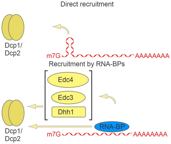
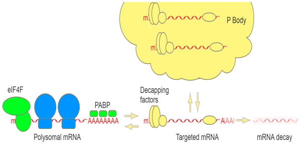
The regulation of mRNA degradation is critical for proper gene expression. Many major pathways for mRNA decay involve the removal of the 5' 7-methyl guanosine (m(7)G) cap in the cytoplasm to allow for 5'-to-3' exonucleolytic decay. The most well studied and conserved eukaryotic decapping enzyme is Dcp2, and its function is aided by co-factors and decapping enhancers. A subset of these factors can act to enhance the catalytic activity of Dcp2, while others might stimulate the remodeling of proteins bound to the mRNA substrate that may otherwise inhibit decapping. Structural studies have provided major insights into the mechanisms by which Dcp2 and decapping co-factors activate decapping. Additional mRNA decay factors can function by recruiting components of the decapping machinery to target mRNAs. mRNA decay factors, decapping factors, and mRNA substrates can be found in cytoplasmic foci named P bodies that are conserved in eukaryotes, though their function remains unknown. In addition to Dcp2, other decapping enzymes have been identified, which may serve to supplement the function of Dcp2 or act in independent decay or quality control pathways. This article is part of a Special Issue entitled: RNA Decay mechanisms.


Figures

Figure 1
Figure 1. The crystal structures of Dcp1 and Dcp2 proteins
(A) Schematic diagrams of the domain organization of Dcp1 and Dcp2 proteins. (B) The crystal structure of the S. pombe Dcp2 (residues 1–266) in the apo form. (C) Crystal structure of S. cerevisiae Dcp1 (green) with the patch 1 region marked. (D) Crystal structure of the EVH1 domain of human Dcp1a (green) in complex with the PNRC2 peptide (pink). (E) and (F) The open and closed conformations of the Dcp1-Dcp2 complex with Dcp1 in green, Dcp2 NTD in orange, Nudix domain in cyan and Nudix motif in red.
Figure 2
Figure 2. The structures of the enhancers of decapping Edc3, Dhh1 and Pat1
(A) Schematic diagrams showing the domain organization of Edc3, Dhh1 and Pat. (B) The crystal structure of YjeF-N dimerization domain from human Edc3 with each subunit colored in pink and deep olive, respectively. (C) The solution structure of the Lsm domain of Edc3 (gray) in complex with the helical leucine-rich motif (HLM) of Dcp2 (cyan). (D) The crystal structure of yeast Dhh1 showing two Rec-A like domains with the N-terminal domain in brown and the C-terminal domain in light blue. (E) The crystal structure of DDX6 C-terminal Rec-A like domain (the ortholog of yeast Dhh1, shown in light blue) in complex with the FDF motif of Edc3 (magenta). (F) The crystal structure of human Pat1 C-terminal α-α superhelical domain.
Figure 3
Figure 3. General principles of decapping complex recruitment
RNA cis-elements, such as the hairpin structure in Rrp41 mRNA, can recruit Dcp2 directly. RNA binding proteins (RNA-BPs) can stimulate decapping by recruiting the Dcp1-Dcp2 complex directly or through decapping enhancers.
Figure 4
Figure 4. P body dynamics
P bodies assemble from translationally repressed, ribosome-free, mRNPs associated with decapping factors. mRNPs assembled into P bodies are either targets of mRNA decay, or can return to translation.

References

    1. Mitchell SF, Walker SE, Algire MA, Park EH, Hinnebusch AG, Lorsch JR. The 5′-7-methylguanosine cap on eukaryotic mRNAs serves both to stimulate canonical translation initiation and to block an alternative pathway. Mol Cell. 2010;39:950–62. - PMC - PubMed
    1. Lewis JD, Izaurralde E. The role of the cap structure in RNA processing and nuclear export. Eur J Biochem. 1997;247:461–9. - PubMed
    1. McKendrick L, Pain VM, Morley SJ. Translation initiation factor 4E. Int J Biochem Cell Biol. 1999;31:31–5. - PubMed
    1. Stevens A. An exoribonuclease from Saccharomyces cerevisiae: effect of modifications of 5′ end groups on the hydrolysis of substrates to 5′ mononucleotides. Biochem Biophys Res Commun. 1978;81:656–61. - PubMed
    1. Li Y, Song M-G, Kiledjian M. Transcript-specific decapping and regulated stability by the human Dcp2 decapping protein. Mol Cell Biol. 2008;28:939–48. - PMC - PubMed