Review
. 2013 Jan 4;112(1):195-208.
doi: 10.1161/CIRCRESAHA.112.266882.

Membrane-associated matrix proteolysis and heart failure

Affiliations
Review

Membrane-associated matrix proteolysis and heart failure

Francis G Spinale et al. Circ Res. .

Abstract

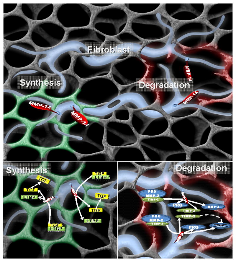
The extracellular matrix (ECM) is a complex entity containing a large portfolio of structural proteins, signaling molecules, and proteases. Changes in the overall integrity and activational state of these ECM constituents can contribute to tissue structure and function, which is certainly true of the myocardium. Changes in the expression patterns and activational states of a family of ECM proteolytic enzymes, the matrix metalloproteinases (MMPs), have been identified in all forms of left ventricle remodeling and can be a contributory factor in the progression to heart failure. However, new clinical and basic research has identified some surprising and unpredicted changes in MMP profiles in left ventricle remodeling processes, such as with pressure or volume overload, as well as with myocardial infarction. From these studies, it has become recognized that proteolytic processing of signaling molecules by certain MMP types, particularly the transmembrane MMPs, actually may facilitate ECM accumulation and modulate fibroblast transdifferentiation; both are critical events in adverse left ventricle remodeling. Based on the ever-increasing substrates and diversity of biological actions of MMPs, it is likely that continued research about the relationship of left ventricle remodeling in this family of proteases will yield new insights into the ECM remodeling process and new therapeutic targets.


Figures

Figure 1
Figure 1
Representative LV myocardial sections taken from remote, border, and MI regions at 14 days post-MI in adult pigs, using methods described previously., (TOP) Immunofluorescent images of these 3 regions following staining with an MMP-14 antisera (Cy3-red). As reported previously in both animal and human LV specimens,,, robust expression of MMP-14 can be observed in both the myocyte and non-myocyte (fibroblast) cell populations. (BOTTOM) Multiple fluorescent labeling was performed whereby nuclear staining was performed using DAPI (blue), phalloidin for actin (green), and a specific antisera for collagen type I (yellow) was performed. Co-localization of MMP-14 will appear as yellow regions. Within the remote region, significant MMP-14 localization can be observed within myocytes and the interstitial space consistent with the transmembrane nature of this MMP type. Within the border region, localization can be appreciated in both viable myocardial cells and proliferating fibroblasts (upper part of panel). Within the MI region, phalloidin positive cells, consistent with myofibroblasts, predominated and co-localized with MMP-14. (Images obtained using a BioRad MRC1024 Confocal Scanning Laser Microscope by Dr. Robert Price, Instrumentation Resource Facility, USC School of Medicine). Scale bar = 30 microns; MMP-14 antisera: Abcam, ab3897, Collagen I antsera: Santa Cruz sc-87048, both used at 1:100 dilution.
Figure 2
Figure 2
(TOP) Schematic of the fibroblast (blue) and ECM in terms of the functional diversity of MMPs; in this case, the representative membrane type MMP, MMP-14. MMP-14 is a transmembrane MMP with a short cytoplasmic tail that likely holds significance for intracellular signaling and potential regulation. The extracellular domain of MMP-14 can cause localized proteolysis of a wide portfolio of ECM proteins, cause a loss of normal ECM-integrin engagement, and activate other MMPs. Thus, MMP-14 can cause a robust and localized amplification of ECM degradation, and in turn, instability. On the other hand, MMP-14 can process profibrotic signaling molecules, such as the release of active TGF, and in turn promote increased fibrillar collagen synthesis and accumulation. In the context of cancer,,, activated cells demonstrate a high degree of MMP polarization and would suggest that these diverse proteolytic actions of MMP-14 can be occurring simultaneously within different ECM locations. With adverse ECM remodeling, such as that with pressure or volume overload or following MI, these ECM degradation and synthesis events can occur concomitantly and be polarized to different subcellular locations as well as to different regions of the LV. Thus, depending upon the context and substrate, MMP-14 can facilitate a loss of normal ECM and replacement fibrosis (such as with pressure overload), a loss of normal ECM and structural support (such as with volume overload), or a combination of both of these proteolytic events (such as with MI). (LOWER LEFT) While MMPs, such as MMP-14, were considered to strictly cause ECM proteolysis, MMP-14 can directly induce a profibrotic cascade involving TGF. MMP-14 can cause proteolysis of the latency binding protein-1 (LTBP-1), which holds TGF in an inactive state, and thereby directly induce a TGF mediated profibrotic signaling cascade. (LOWER RIGHT) Another critical function of MMP-14 is the complex formation with pro-MMP-2 and TIMP-2, which will result in an active form of MMP-2. Moreover, TIMP-2 can then bind to the active site of MMP-2, which will extinguish proteolytic activity. This localized activation-inhibition cycle can provide for precise ECM proteolysis. Thus, the type and location of the MMP-14 substrate very likely dictates the effects upon ECM structure and function. (Initial image provided courtesy of Shaun Riffle, USC School of Medicine, and annotation by Craig P. Novack).
Figure 3
Figure 3
Dual isotope hybrid SPECT/CT imaging obtained using Thallium-201 (201Tl) and a technetium-99m labeled MMP targeted tracer (99mTc-RP805) in a canine model of MI induced by balloon coronary occlusion. The imaging approaches and validation have been reported previously. LV myocardial perfusion by 201TI is designated as green, and MMP radiotracer uptake is designated as red. Significant MMP radiotracer uptake could be observed within the LV myocardium, indicative of MMP proteolytic activity, at 3 and 14 days post-MI. Moreover, MMP activity appears to occur within normally perfused LV regions at later post-MI time points. (Images courtesy of Dr. Albert Sinusas, Professor of Medicine and Diagnostic Radiology Director, Cardiovascular Imaging Director, Yale Translational Research Imaging Center, Yale University School of Medicine).

References

    1. Konstam MA, Kramer DG, Patel AR, Maron MS, Udelson JE. Left ventricular remodeling in heart failure: current concepts in clinical significance and assessment. JACC Cardiovasc Imaging. 2011 Jan;4(1):98–108. - PubMed
    1. Quiñones MA, Greenberg BH, Kopelen HA, Koilpillai C, Limacher MC, Shindler DM, Shelton BJ, Weiner DH. Echocardiographic predictors of clinical outcome in patients with left ventricular dysfunction enrolled in the SOLVD registry and trials: significance of left ventricular hypertrophy. Studies of Left Ventricular Dysfunction. J Am Coll Cardiol. 2000 Apr;35(5):1237–44. - PubMed
    1. White HD, Norris RM, Brown MA, Brandt PW, Whitlock RM, Wild CJ. Left ventricular end-systolic volume as the major determinant of survival after recovery from myocardial infarction. Circulation. 1987 Jul;76(1):44–51. - PubMed
    1. Weir RA, McMurray JJ, Velazquez EJ. Epidemiology of heart failure and left ventricular systolic dysfunction after acute myocardial infarction: prevalence, clinical characteristics, and prognostic importance. Am J Cardiol. 2006 May 22;97(10A):13F–25F. Epub 2006 Apr 21. - PubMed
    1. Sutton MG, Sharpe N. Left ventricular remodeling after myocardial infarction: pathophysiology and therapy. Circulation. 2000 Jun 27;101(25):2981–8. - PubMed

Publication types

MeSH terms

Substances