Review
. 2013 Mar-Apr;7(2):174-86.
doi: 10.4161/cam.23130. Epub 2013 Jan 3.

Membrane tubulovesicular extensions (cytonemes): secretory and adhesive cellular organelles

Affiliations
Review

Membrane tubulovesicular extensions (cytonemes): secretory and adhesive cellular organelles

Svetlana I Galkina et al. Cell Adh Migr. 2013 Mar-Apr.

Abstract

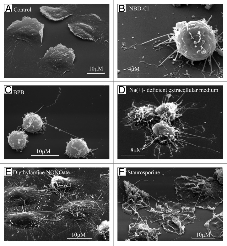
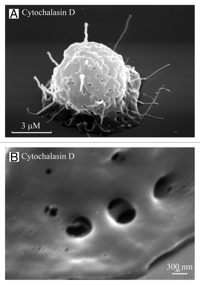
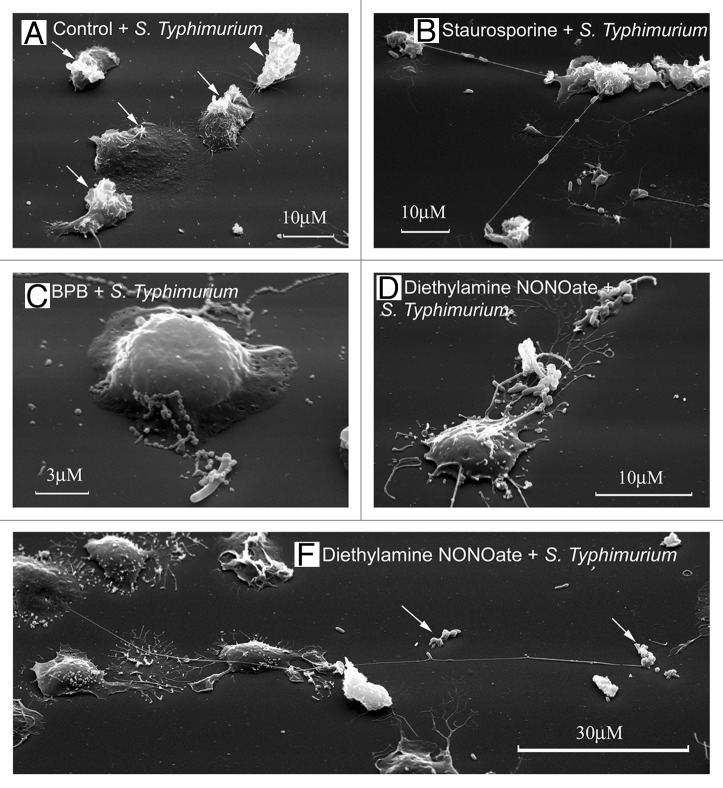
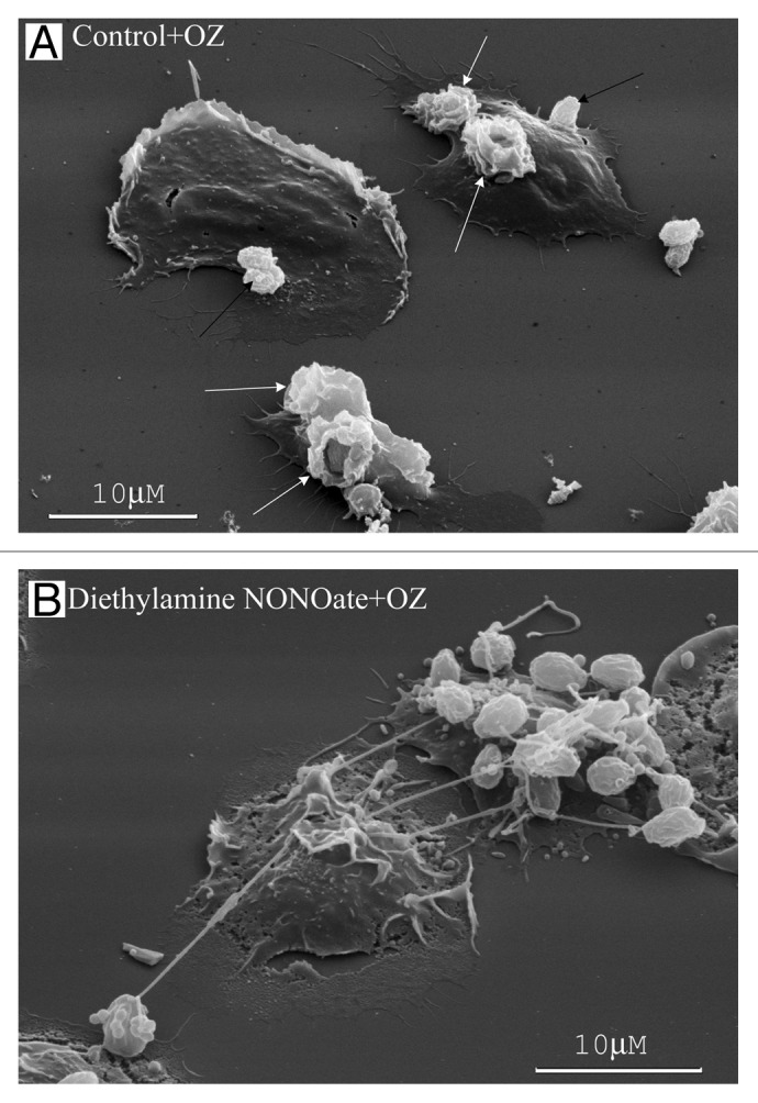
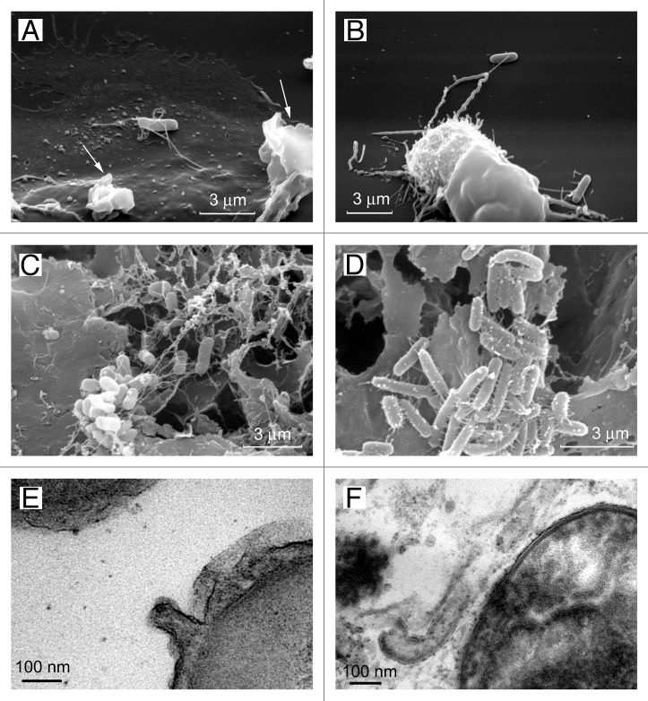
In this review, we summarized data on the formation and structure of the long and highly adhesive membrane tubulovesicular extensions (TVEs, membrane tethers or cytonemes) observed in human neutrophils and other mammalian cells, protozoan parasites and bacteria. We determined that TVEs are membrane protrusions characterized by a uniform diameter (130-250 nm for eukaryotic cells and 60-90 nm for bacteria) along the entire length, an outstanding length and high rate of development and a high degree of flexibility and capacity for shedding from the cells. This review represents TVEs as protrusions of the cellular secretory process, serving as intercellular adhesive organelles in eukaryotic cells and bacteria. An analysis of the physical and chemical approaches to induce TVEs formation revealed that disrupting the actin cytoskeleton and inhibiting glucose metabolism or vacuolar-type ATPase induces TVE formation in eukaryotic cells. Nitric oxide is represented as a physiological regulator of TVE formation.

Keywords: bacteria; cell secretion; cell-cell communications; cytonemes; eukaryotic cells; exocytotic carriers; host-pathogen interactions; membrane tethers; membrane tubulovesicular extensions (TVEs); protozoan parasites.


Figures

None
Figure 1. Formation of TVE in human neutrophils upon adhesion to the fibronectin-coated substrata. Scanning electron microscopy images of human neutrophils plated onto fibronectin-coated substrata for 20 min at 37°C: (A) in control conditions; (B) in the presence of inhibitor of V-ATPase NBD-Cl (100 µm); (C) in the presence of BPB (10 μM); (D) in the extracellular medium, where Na+ ions were substituted with K+ ions; (E) in the presence of the NO donor diethylamine NONOate (1 mM) or (F) microbial alkaloid staurosporine (200 nM).,-
None
Figure 2. TVEs and invaginations on neutrophil bodies. Scanning electron images of neutrophils plated onto fibronectin in the presence of the actin-disrupting agent cytochalasin D (10 µg/ml) for 20 (A) and 40 (B) min. TVEs and specific invaginations were observed on the cell body. (B) The invaginations on the neutrophil bodies resemble “porosomes” of exocrine and neuroendocrine cells, with two depressions.
None
Figure 3. Neutrophil TVEs bind and hold pathogenic bacteria Salmonella enterica serovar Typhimurium over long distances. Scanning electron microscopy images of human neutrophils plated onto fibronectin-coated substrata for 20 min at 37°C: (A) in the control conditions, (B) in the presence of staurosporine (200 nM), (C) in the presence of BPB (10 μM) or (E) in the presence of the NO donor diethylamine NONOate (1 mM). At the end of incubation, serum-opsonized S. Typhimurium bacteria (bacteria/cells ratio 20:1) were added to neutrophils for 5 min. The neutrophils plated at control conditions phagocytosed the bacteria. “Ruffles” formed on the cell surface where bacteria entered the cell [(A), arrows]. The neutrophils plated onto fibronectin in the presence of inducers of TVE formation (B–E) bound and held bacteria through TVEs.,
None
Figure 4. NO alters human neutrophil interactions with yeast. (A) The neutrophils plated onto fibronectin under the control conditions phagocytosed opsonized zymosan particles (dried yeast coated with serum) forming specific “cups” (white arrows) on the cell surface or bound yeast particles onto the cell surface (black arrows). (B) The neutrophils plated onto fibronectin in the presence of the NO donor diethylamine NONOate (1 mM) bound opsonized zymosan particles through TVEs.
None
Figure 5. Cytonemes of bacteria. The scanning electron microscopy images show Salmonella enterica serovar Typhimurium attached to the surface of the control neutrophils (A) or to the TVEs of BPB-treated neutrophils (B) through cytonemes. Salmonella of the virulent C53 strain (C) and the non-flagellated SJW880 strain (D) were interconnected through cytonemes in biofilms grown on the surface of gallstones. Transmission electron microscopy images of 60-nm membrane tubules derived from the outer membrane of the bacteria (E and F).

Similar articles

Cited by

References

    1. Gustafson T, Wolpert L. Cellular movement and contact in sea urchin morphogenesis. Biol Rev Camb Philos Soc. 1967;42:442–98. doi: 10.1111/j.1469-185X.1967.tb01482.x. - DOI - PubMed
    1. Solursh M, Lane MC. Extracellular matrix triggers a directed cell migratory response in sea urchin primary mesenchyme cells. Dev Biol. 1988;130:397–401. doi: 10.1016/0012-1606(88)90445-9. - DOI - PubMed
    1. Miller J, Fraser SE, McClay D. Dynamics of thin filopodia during sea urchin gastrulation. Development. 1995;121:2501–11. - PubMed
    1. Ramírez-Weber FA, Kornberg TB. Cytonemes: cellular processes that project to the principal signaling center in Drosophila imaginal discs. Cell. 1999;97:599–607. - PubMed
    1. Shao JY, Hochmuth RM. Micropipette suction for measuring piconewton forces of adhesion and tether formation from neutrophil membranes. Biophys J. 1996;71:2892–901. doi: 10.1016/S0006-3495(96)79486-9. - DOI - PMC - PubMed

Publication types

LinkOut - more resources