Arabinoxylan rice bran (MGN-3/Biobran) provides protection against whole-body γ-irradiation in mice via restoration of hematopoietic tissues
- PMID: 23287771
- PMCID: PMC3650744
- DOI: 10.1093/jrr/rrs119
Arabinoxylan rice bran (MGN-3/Biobran) provides protection against whole-body γ-irradiation in mice via restoration of hematopoietic tissues
Abstract
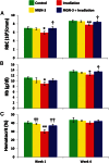
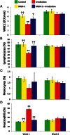
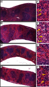
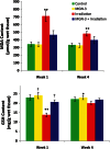
The aim of the current study is to examine the protective effect of MGN-3 on overall maintenance of hematopoietic tissue after γ-irradiation. MGN-3 is an arabinoxylan from rice bran that has been shown to be a powerful antioxidant and immune modulator. Swiss albino mice were treated with MGN-3 prior to irradiation and continued to receive MGN-3 for 1 or 4 weeks. Results were compared with mice that received radiation (5 Gy γ rays) only, MGN-3 (40 mg/kg) only and control mice (receiving neither radiation nor MGN-3). At 1 and 4 weeks post-irradiation, different hematological, histopathological and biochemical parameters were examined. Mice exposed to irradiation alone showed significant depression in their complete blood count (CBC) except for neutrophilia. Additionally, histopathological studies showed hypocellularity of their bone marrow, as well as a remarkable decrease in splenic weight/relative size and in number of megakaryocytes. In contrast, pre-treatment with MGN-3 resulted in protection against irradiation-induced damage to the CBC parameters associated with complete bone marrow cellularity, as well as protection of the aforementioned splenic changes. Furthermore, MGN-3 exerted antioxidative activity in whole-body irradiated mice, and provided protection from irradiation-induced loss of body and organ weight. In conclusion, MGN-3 has the potential to protect progenitor cells in the bone marrow, which suggests the possible use of MGN-3/Biobran as an adjuvant treatment to counteract the severe adverse side effects associated with radiation therapy.
Keywords: Biobran; MGN-3; hematopoeitic cells; radiation.
Figures


Similar articles
-
Arabinoxylan rice bran (MGN-3/Biobran) enhances radiotherapy in animals bearing Ehrlich ascites carcinoma†.J Radiat Res. 2019 Nov 22;60(6):747-758. doi: 10.1093/jrr/rrz055. J Radiat Res. 2019. PMID: 31504707 Free PMC article.
-
Protection of Swiss albino mice against whole-body gamma irradiation by diltiazem.Br J Radiol. 2007 Feb;80(950):77-84. doi: 10.1259/bjr/41714035. Epub 2006 Oct 26. Br J Radiol. 2007. PMID: 17068014
-
Arabinoxylan rice bran (MGN-3/Biobran) alleviates radiation-induced intestinal barrier dysfunction of mice in a mitochondrion-dependent manner.Biomed Pharmacother. 2020 Apr;124:109855. doi: 10.1016/j.biopha.2020.109855. Epub 2020 Jan 24. Biomed Pharmacother. 2020. PMID: 31986410
-
Evidence-Based Review of BioBran/MGN-3 Arabinoxylan Compound as a Complementary Therapy for Conventional Cancer Treatment.Integr Cancer Ther. 2018 Jun;17(2):165-178. doi: 10.1177/1534735417735379. Epub 2017 Oct 17. Integr Cancer Ther. 2018. PMID: 29037071 Free PMC article. Review.
-
A contribution to the study of damage and regeneration of hemopoiesis during fractionated irradiation and repeated bone marrow transplantation.Strahlenther Onkol. 1988 Jun;164(6):357-62. Strahlenther Onkol. 1988. PMID: 3291165 Review.
Cited by
-
Modified Rice Bran Arabinoxylan by Lentinus edodes Mycelial Enzyme as an Immunoceutical for Health and Aging-A Comprehensive Literature Review.Molecules. 2023 Aug 29;28(17):6313. doi: 10.3390/molecules28176313. Molecules. 2023. PMID: 37687141 Free PMC article. Review.
-
Protective effect of hydroferrate fluid, MRN-100, against lethality and hematopoietic tissue damage in γ-radiated Nile tilapia, Oreochromis niloticus.J Radiat Res. 2013 Sep;54(5):852-62. doi: 10.1093/jrr/rrt029. Epub 2013 Apr 14. J Radiat Res. 2013. PMID: 23589025 Free PMC article.
-
Protective Effect of Biobran/MGN-3 against Sporadic Alzheimer's Disease Mouse Model: Possible Role of Oxidative Stress and Apoptotic Pathways.Oxid Med Cell Longev. 2021 Jan 26;2021:8845064. doi: 10.1155/2021/8845064. eCollection 2021. Oxid Med Cell Longev. 2021. PMID: 33574982 Free PMC article.
-
Melanin nanoparticles (MNPs) provide protection against whole-body ɣ-irradiation in mice via restoration of hematopoietic tissues.Mol Cell Biochem. 2015 Jan;399(1-2):59-69. doi: 10.1007/s11010-014-2232-y. Epub 2014 Oct 10. Mol Cell Biochem. 2015. PMID: 25300618
-
The Effect of a Hydrolyzed Polysaccharide Dietary Supplement on Biomarkers in Adults with Nonalcoholic Fatty Liver Disease.Evid Based Complement Alternat Med. 2018 May 3;2018:1751583. doi: 10.1155/2018/1751583. eCollection 2018. Evid Based Complement Alternat Med. 2018. PMID: 29853945 Free PMC article.
References
-
- Badr El-Din NK. Protective role of sanumgerman against γ-irradiation-induced oxidative stress in Ehrlich carcinoma-bearing mice. Nutr Res. 2004;24:271–91.
-
- Karbownik M, Reiter RJ. Antioxidative effects of melatonin in protection against cellular damage caused by ionizing radiation. Proc Soc Exp Biol Med. 2000;225:9–22. - PubMed
-
- Karran P. DNA double strand break repair in mammalian cells. Curr Opin Genet Dev. 2000;10:144–50. - PubMed
-
- Kalpana KB, Devipriya N, Srinivasan M, et al. Evaluating the radioprotective effect of hesperidin in the liver of Swiss albino mice. Eur J Pharmacol. 2011;658:6–12. - PubMed
Publication types
MeSH terms
Substances
LinkOut - more resources
Full Text Sources
Other Literature Sources
Medical

