

Quantitative analysis of murine terminal erythroid differentiation in vivo: novel method to study normal and disordered erythropoiesis
- PMID: 23287863
- PMCID: PMC3578961
- DOI: 10.1182/blood-2012-09-456079
Quantitative analysis of murine terminal erythroid differentiation in vivo: novel method to study normal and disordered erythropoiesis
Abstract
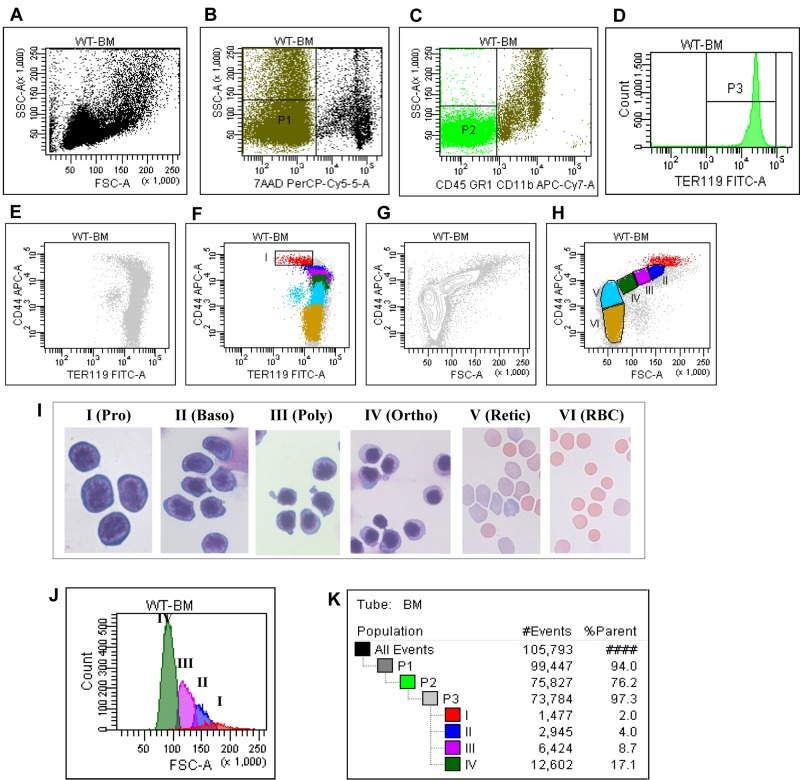
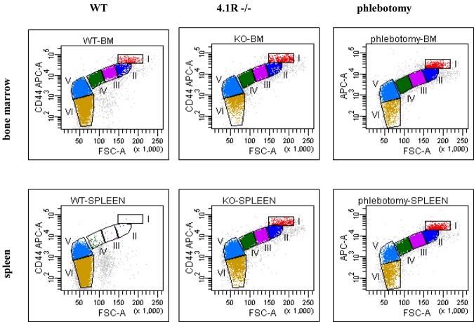
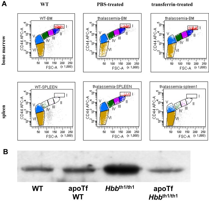
Terminal erythroid differentiation is the process during which proerythroblasts differentiate to produce enucleated reticulocytes. Although it is well established that during murine erythropoiesis in vivo, 1 proerythroblast undergoes 3 mitosis to generate sequentially 2 basophilic, 4 polychromatic, and 8 orthochromatic erythroblasts, currently there is no method to quantitatively monitor this highly regulated process. Here we outline a method that distinguishes each distinct stage of erythroid differentiation in cells from mouse bone marrow and spleen based on expression levels of TER119, CD44, and cell size. Quantitative analysis revealed that the ratio of proerythroblasts:basophilic:polychromatic:orthromatic erythroblasts follows the expected 1:2:4:8 ratio, reflecting the physiologic progression of terminal erythroid differentiation in normal mice. Moreover, in 2 stress erythropoiesis mouse models, phlebotomy-induced acute anemia and chronic hemolytic anemia because of 4.1R deficiency, the ratio of these erythroblast populations remains the same as that of wild-type bone marrow. In contrast, in anemic β-thalassemia intermedia mice, there is altered progression which is restored to normal by transferrin treatment which was previously shown to ameliorate the anemic phenotype. The means to quantitate in vivo murine erythropoiesis using our approach will probably have broad application in the study of altered erythropoiesis in various red cell disorders.
Figures
References
-
- McLeod DL, Shreeve MM, Axelrad AA. Improved plasma culture system for production of erythrocytic colonies in vitro: quantitative assay method for CFU-E. Blood. 1974;44(4):517–534. - PubMed
-
- Heath DS, Axelrad AA, McLeod DL, Shreeve MM. Separation of the erythropoietin-responsive progenitors BFU-E and CFU-E in mouse bone marrow by unit gravity sedimentation. Blood. 1976;47(5):777–792. - PubMed
-
- Dover GJ, Chan T, Sieber F. Fetal hemoglobin production in cultures of primitive and mature human erythroid progenitors: differentiation affects the quantity of fetal hemoglobin produced per fetal-hemoglobin-containing cell. Blood. 1983;61(6):1242–1246. - PubMed
-
- Socolovsky M, Nam H, Fleming MD, Haase VH, Brugnara C, Lodish HF. Ineffective erythropoiesis in Stat5a(−/−)5b(−/−) mice due to decreased survival of early erythroblasts. Blood. 2001;98(12):3261–3273. - PubMed
Publication types
MeSH terms
Substances
Grants and funding
LinkOut - more resources
Full Text Sources
Other Literature Sources
Molecular Biology Databases
Miscellaneous
