

A flexible annular-array imaging platform for micro-ultrasound
- PMID: 23287923
- PMCID: PMC3738186
- DOI: 10.1109/TUFFC.2013.2548
A flexible annular-array imaging platform for micro-ultrasound
Abstract
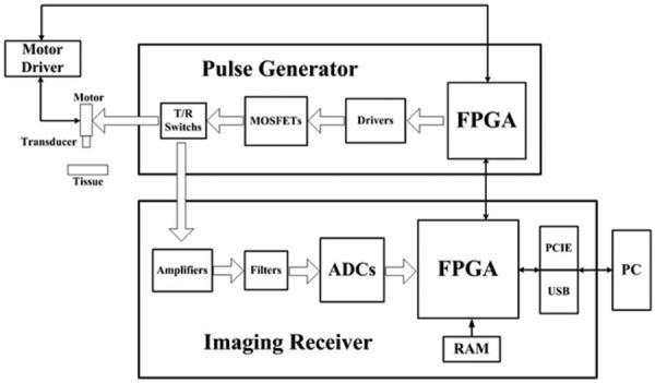
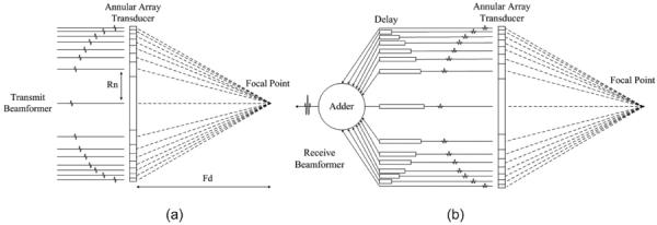
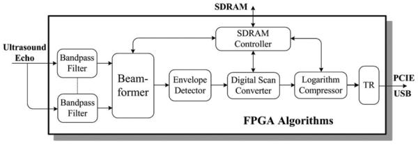
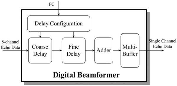
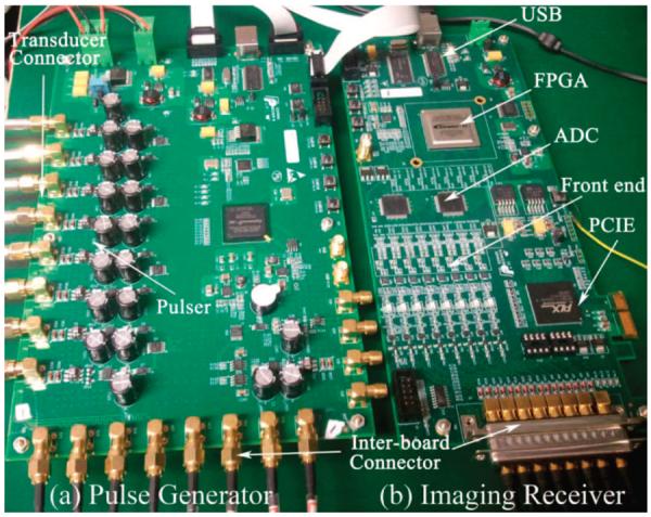
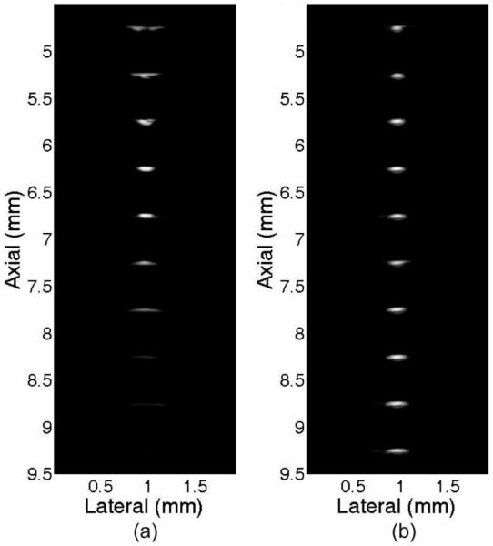
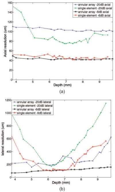
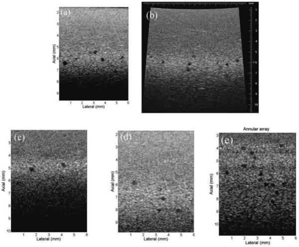
Micro-ultrasound is an invaluable imaging tool for many clinical and preclinical applications requiring high resolution (approximately several tens of micrometers). Imaging systems for micro-ultrasound, including single-element imaging systems and linear-array imaging systems, have been developed extensively in recent years. Single-element systems are cheaper, but linear-array systems give much better image quality at a higher expense. Annular-array-based systems provide a third alternative, striking a balance between image quality and expense. This paper presents the development of a novel programmable and real-time annular-array imaging platform for micro-ultrasound. It supports multi-channel dynamic beamforming techniques for large-depth-of-field imaging. The major image processing algorithms were achieved by a novel field-programmable gate array technology for high speed and flexibility. Real-time imaging was achieved by fast processing algorithms and high-speed data transfer interface. The platform utilizes a printed circuit board scheme incorporating state-of-the-art electronics for compactness and cost effectiveness. Extensive tests including hardware, algorithms, wire phantom, and tissue mimicking phantom measurements were conducted to demonstrate good performance of the platform. The calculated contrast-to-noise ratio (CNR) of the tissue phantom measurements were higher than 1.2 in the range of 3.8 to 8.7 mm imaging depth. The platform supported more than 25 images per second for real-time image acquisition. The depth-of-field had about 2.5-fold improvement compared to single-element transducer imaging.
Figures


References
-
- Foster FS, Pavlin CJ, Harasiewicz KA, Christopher DA, Turnbull DH. Advances in ultrasound biomicroscopy. Ultrasound Med. Biol. 2000;26(no. 1):1–27. - PubMed
-
- Gazzard G, Friedman DS, Devereux JG, Chew P, Seah SK. A prospective ultrasound biomicroscopy evaluation of changes in anterior segment morphology after laser iridotomy in Asian eyes. Ophthalmology. 2003;110(no. 3):630–638. - PubMed
-
- Huang Y, Zheng Y, Leung SF, Choi AP. High frequency ultrasound assessment of skin fibrosis: Clinical results. Ultrasound Med. Biol. 2007;33(no. 8):1191–1198. - PubMed
-
- Vogt M, Ermert H. In vivo ultrasound biomicroscopy of skin: Spectral system characteristics and inverse filtering optimization. IEEE Trans. Ultrason. Ferroelectr. Freq. Control. 2007;54(no. 8):1551–1559. - PubMed
-
- de Korte CL, van der Steen AFW, Cespedes EI, Pasterkamp G, Carlier SG, Mastik F, Schoneveld AH, Serruys PW, Bom N. Characterization of plaque components and vulnerability with intravascular ultrasound elastography. Phys. Med. Biol. 2000;45(no. 6):1465–1475. - PubMed
Publication types
MeSH terms
Grants and funding
LinkOut - more resources
Full Text Sources
Other Literature Sources
