

Dp44mT targets the AKT, TGF-β and ERK pathways via the metastasis suppressor NDRG1 in normal prostate epithelial cells and prostate cancer cells
- PMID: 23287991
- PMCID: PMC3566801
- DOI: 10.1038/bjc.2012.582
Dp44mT targets the AKT, TGF-β and ERK pathways via the metastasis suppressor NDRG1 in normal prostate epithelial cells and prostate cancer cells
Erratum in
-
Correction to: Dp44mT targets the AKT, TGF-β and ERK pathways via the metastasis suppressor NDRG1 in normal prostate epithelial cells and prostate cancer cells.Br J Cancer. 2026 Apr;134(7):1116-1120. doi: 10.1038/s41416-026-03353-w. Br J Cancer. 2026. PMID: 41772277 Free PMC article. No abstract available.
Abstract
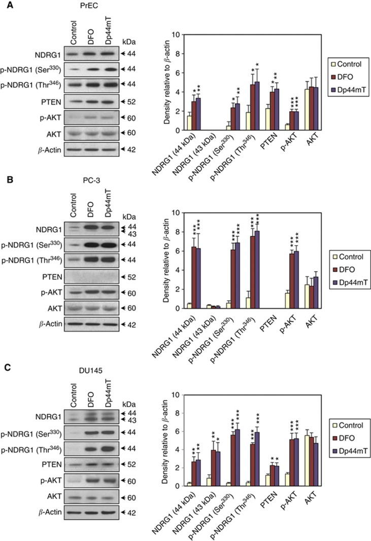
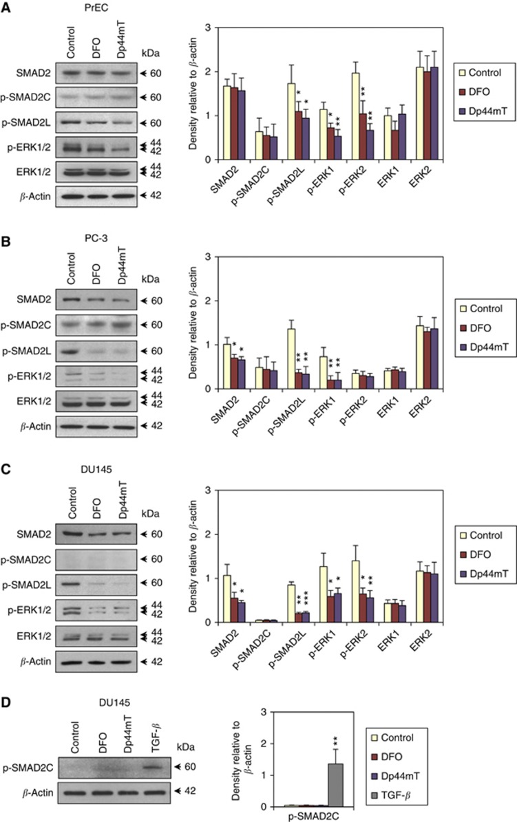
Background: Effective treatment of prostate cancer should be based on targeting interactions between tumour cell signalling pathways and key converging downstream effectors. Here, we determined how the tumourigenic phosphoinositide 3-kinase/protein kinase B (PI3K/AKT), tumour-suppressive phosphatase and tensin homologue deleted on chromosome 10 (PTEN) and transforming growth factor-β (TGF-β) pathways are integrated via the metastasis suppressor, N-myc downstream-regulated gene-1 (NDRG1). Moreover, we assessed how the novel anti-tumour agent, Dp44mT, may target these integrated pathways by increasing NDRG1 expression.
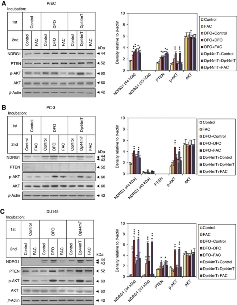
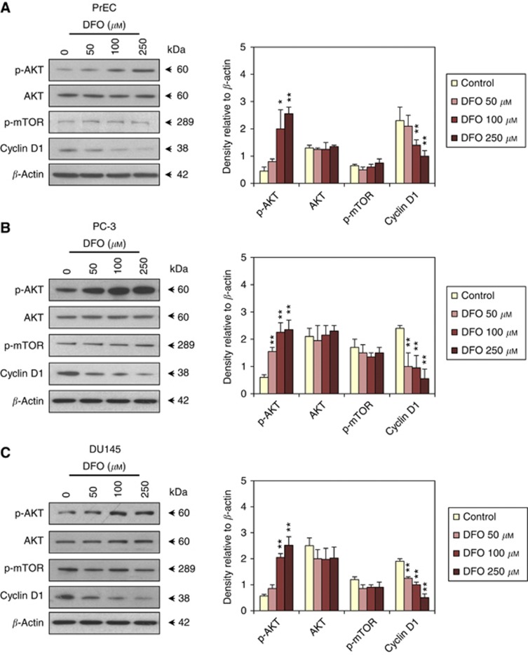
Methods: Protein expression in Dp44mT-treated normal human prostate epithelial cells and prostate cancer cells (PC-3, DU145) was assessed by western blotting. The role of NDRG1 was examined by transfection using an NDRG1 overexpression vector or shRNA.
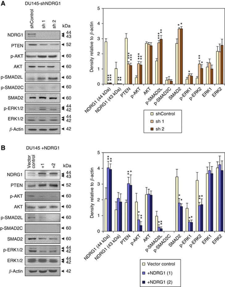
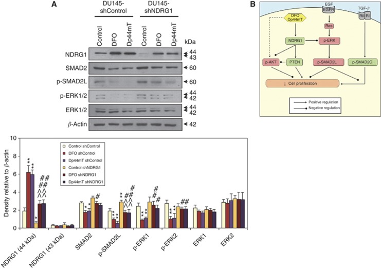
Results: Dp44mT increased levels of tumour-suppressive PTEN, and decreased phosphorylation of ERK1/2 and SMAD2L, which are regulated by oncogenic Ras/MAPK signalling. Importantly, the effects of Dp44mT on NDRG1 and p-SMAD2L expression were more marked in prostate cancer cells than normal prostate epithelial cells. This may partly explain the anti-tumour selectivity of these agents. Silencing NDRG1 expression increased phosphorylation of tumourigenic AKT, ERK1/2 and SMAD2L and decreased PTEN levels, whereas NDRG1 overexpression induced the opposite effect. Furthermore, NDRG1 silencing significantly reduced the ability of Dp44mT to suppress p-SMAD2L and p-ERK1/2 levels.
Conclusion: NDRG1 has an important role in mediating the tumour-suppressive effects of Dp44mT in prostate cancer via selective targeting of the PI3K/AKT, TGF-β and ERK pathways.
Figures
References
-
- Alvarez-Tejado M, Naranjo-Suarez S, Jimenez C, Carrera AC, Landazuri MO, del Peso L. Hypoxia induces the activation of the phosphatidylinositol 3-kinase/Akt cell survival pathway in PC12 cells - Protective role in apoptosis. J Biol Chem. 2001;276 (25:22368–22374. - PubMed
-
- Assinder SJ, Dong Q, Kovacevic Z, Richardson DR. The TGF-beta, PI3K/Akt and PTEN pathways: established and proposed biochemical integration in prostate cancer. Biochem J. 2009;417 (2:411–421. - PubMed
-
- Assinder SJ, Dong Q, Mangs H, Richardson DR. Pharmacological targeting of the integrated AKT, PTEN and TGF-beta pathways in prostate cancer. Mol Pharmacol. 2008;75 (3:429–436. - PubMed
-
- Bandyopadhyay S, Pai SK, Gross SC, Hirota S, Hosobe S, Miura K, Saito K, Commes T, Hayashi S, Watabe M, Watabe K. The Drg-1 gene suppresses tumor metastasis in prostate cancer. Cancer Res. 2003;63 (8:1731–1736. - PubMed
-
- Bandyopadhyay S, Pai SK, Hirota S, Hosobe S, Takano Y, Saito K, Piquemal D, Commes T, Watabe M, Gross SC, Wang Y, Ran S, Watabe K. Role of the putative tumor metastasis suppressor gene Drg-1 in breast cancer progression. Oncogene. 2004a;23 (33:5675–5681. - PubMed
Publication types
MeSH terms
Substances
LinkOut - more resources
Full Text Sources
Other Literature Sources
Medical
Molecular Biology Databases
Research Materials
Miscellaneous
