

Photodynamic therapy for retinal capillary hemangioma
- PMID: 23288135
- PMCID: PMC3597867
- DOI: 10.1038/eye.2012.259
Photodynamic therapy for retinal capillary hemangioma
Abstract
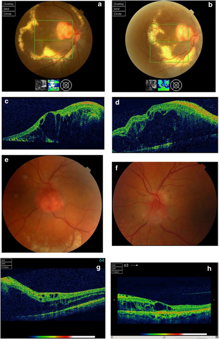
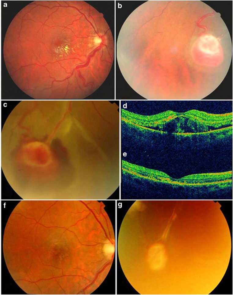
Purpose: To describe the results of photodynamic therapy (PDT) for juxtapapillary and peripheral retinal capillary hemangioma (RCH).
Patients and methods: Interventional case series of four eyes (four patients) with juxtapapillary RCH and one eye (one patient) with peripheral RCH. Two eyes with juxtapapillary RCH had received two sessions of full-fluence, double-duration PDT; whereas other two eyes had received single session of half-fluence, single-duration PDT. The peripheral RCH was treated with a single session of full-fluence, single-duration PDT.
Results: Two patients had von Hippel-Lindau disease. Follow-up duration ranged from 4 months to 1 year. Pre-PDT visual acuity (VA) ranged from 20/200 to HM (juxtapapillary RCH) and 20/100 (peripheral RCH). Among the eyes with juxtapapillary RCH, tumor regression with partial resolution of macular edema was noted in two eyes (one eye each with half-fluence and full-fluence PDT), whereas two eyes had no change in tumor size with persistent macular edema. VA remained stable in three eyes and declined in one eye. In an eye with peripheral RCH, regression of tumor and macular edema with VA improvement was noted. Post-PDT complications included epiretinal membrane (one eye) and transient exudative retinal detachment (one eye).
Conclusion: PDT can be effective in reducing macular edema associated with RCH but this does not always correspond with an improvement in VA especially for juxtapapillary tumors.
Figures
References
-
- Singh AD, Nouri M, Shields CL, Shields JA, Perez N. Treatment of retinal capillary haemangioma. Ophthalmology. 2002;109:1799–1806. - PubMed
-
- Schmidt-Erfurth UM, Kusserow C, Barbazetto IA, Laqua H. Benefits and complications of photodynamic therapy of papillary capillary haemangiomas. Ophthalmology. 2002;109 (7:1256–1266. - PubMed
-
- Atebara NH. Retinal capillary haemangioma treated with verteporfin photodynamic therapy. Am J Ophthalmol. 2002;134:788–790. - PubMed
-
- Aaberg JrTM, Aaberg SrTM, Martin DF, Gilman JP, Myles R. Three cases of large retinal capillary haemangiomas treated with verteporfin and photodynamic therapy. Arch Ophthalmol. 2005;123 (3:328–332. - PubMed
-
- Golshevsky JR, O'Day J. Photodynamic therapy in the management of juxtapapillary capillary haemangiomas. Clin Experiment Ophthalmol. 2005;33 (5:509–512. - PubMed
Publication types
MeSH terms
Substances
LinkOut - more resources
Full Text Sources
Other Literature Sources
