. 2013 Apr;33(4):718-26.
doi: 10.1161/ATVBAHA.112.300329. Epub 2013 Jan 3.

Peptide inhibitor of CXCL4-CCL5 heterodimer formation, MKEY, inhibits experimental aortic aneurysm initiation and progression

Affiliations

Peptide inhibitor of CXCL4-CCL5 heterodimer formation, MKEY, inhibits experimental aortic aneurysm initiation and progression

Yasunori Iida et al. Arterioscler Thromb Vasc Biol. 2013 Apr.

Abstract

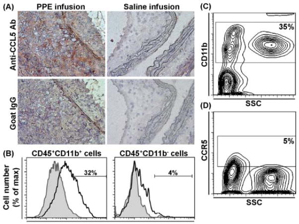
Objective: Macrophages are critical contributors to abdominal aortic aneurysm (AAA) disease. We examined the ability of MKEY, a peptide inhibitor of CXCL4-CCL5 interaction, to influence AAA progression in murine models.

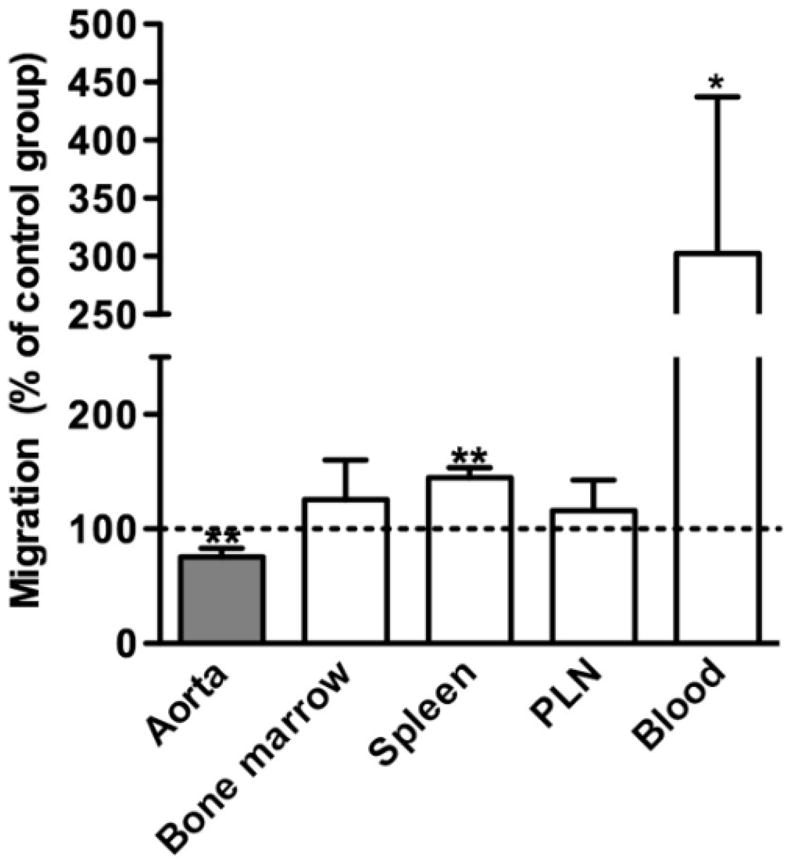
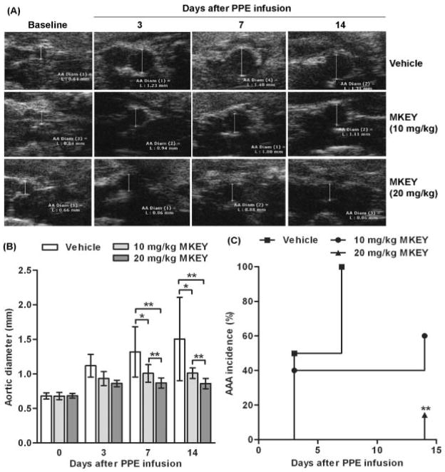
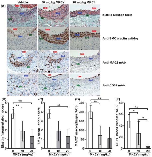
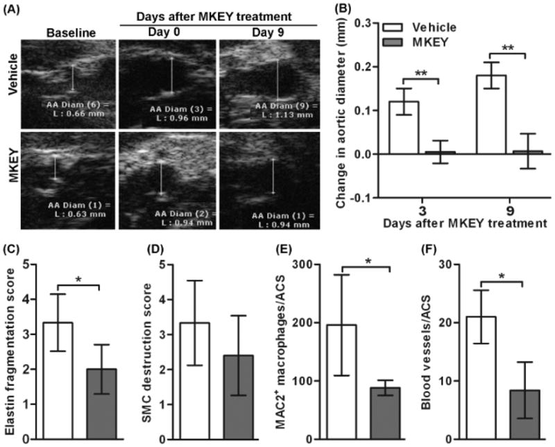
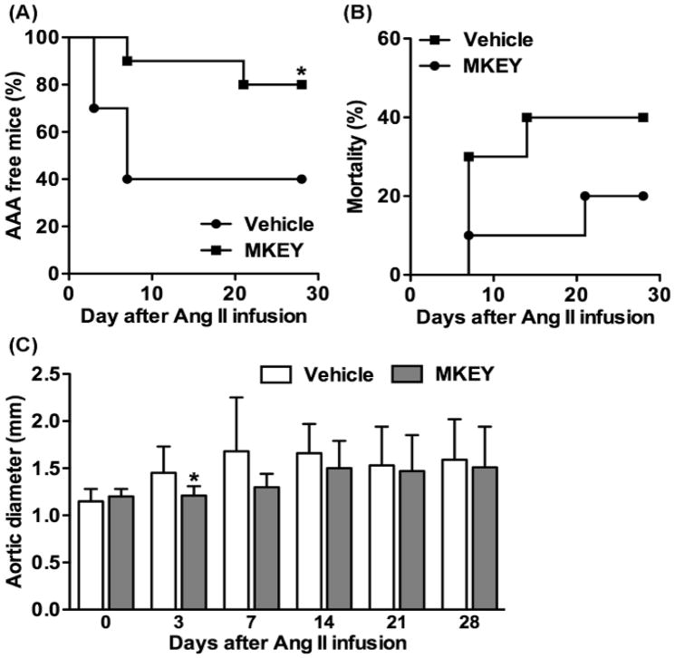
Approach and results: AAAs were created in 10-week-old male C57BL/6J mice by transient infrarenal aortic porcine pancreatic elastase infusion. Mice were treated with MKEY via intravenous injection either (1) before porcine pancreatic elastase infusion or (2) after aneurysm initiation. Immunostaining demonstrated CCL5 and CCR5 expression on aneurysmal aortae and mural monocytes/macrophages, respectively. MKEY treatment partially inhibited migration of adaptively transferred leukocytes into aneurysmal aortae in recipient mice. Although all vehicle-pretreated mice developed AAAs, aneurysms formed in only 60% (3/5) and 14% (1/7) of mice pretreated with MKEY at 10 and 20 mg/kg, respectively. MKEY pretreatment reduced aortic diameter enlargement, preserved medial elastin fibers and smooth muscle cells, and attenuated mural macrophage infiltration, angiogenesis, and aortic metalloproteinase 2 and 9 expression after porcine pancreatic elastase infusion. MKEY initiated after porcine pancreatic elastase infusion also stabilized or reduced enlargement of existing AAAs. Finally, MKEY treatment was effective in limiting AAA formation after angiotensin II infusion in apolipoprotein E-deficient mice.

Conclusions: MKEY suppresses AAA formation and progression in 2 complementary experimental models. Peptide inhibition of CXCL4-CCL5 interactions may represent a viable translational strategy to limit progression of human AAA disease.


Figures

Figure 1
Figure 1. Expression of CCL5 and its leukocyte receptor CCR5 in aneurysmal aortae
(A): Acetone-fixed frozen sections from mice 2 wk after PPE (left panels) or saline (right panels) infusion were stained with a goat anti-mouse CCL5 polyclonal antibody (upper panels) or an equal amount of normal goat IgG (negative control, lower panels). This staining pattern was reproduced at least 3 mice. Original magnification: X400. (B): Single leukocyte suspensions from enzyme-digested aneurysmal aortae were stained with the mAbs against CD45, CD11b and CCR5 (or its negative control antibody), and analyzed using flow cytometry. The percentages of CCR5+ cells in CD45+CD11b+ (monocytes/macrophages) and CD45+CD11b cells (other leukocytes) are shown in the left and right panels, respectively. Each flow cytometric histograph is the overlay image for the staining of anti-CCR5 mAb (unshaded) and its negative control antibody (shaded). This experiment was repeated 3 times, and the cells pooled from 3 mouse aortae were used for each staining. (C, D): Whole blood leukocytes from mice 2 wk after PPE infusion were stained with CD11b and CCR5 (or its negative control antibody), and analyzed using flow cytometry. Both small and large leukocytes expressed CD11b (C). A representative flow cytometric plot shows that 9.4% of CD11b+ leukocytes expressed CCR5 and most of them were small leukocytes as indicated by side scatter size (D). This experiment was repeated in 5 mice.
Figure 2
Figure 2. Leukocyte migration in experimental AAA
A mixed population of spleen and bone marrow cells from aneurysmal mice were labeled with CFSE, and intravenously transferred into aneurysmal recipient mice pretreated with MKEY 30 min before cell transfer. Recipient mice were sacrificed 2 hours after cell transfer. Donor cells in the spleen, peripheral lymph nodes (PLN), bone marrow and aneurysmal aortae of recipient mice were evaluated using either flow cytometric or tissue immunofluorescence analysis. Migration of donor cells in MKEY-treated group was expressed as the percentage of that in vehicle-treated group, in which migration was set up at 100. Nonparametric Mann-Whitney test, *P<0.05 and **P<0.01 compared to vehicle-treated group. n=4 mice in each group.
Figure 3
Figure 3. Influence of MKEY treatment on AAA formation and progression
Male C57BL/6 mice were treated with vehicle (n=8), 10 mg/kg MKEY (n=5) or 20 mg/kg MKEY (n=7) starting on day 3 prior to PE infusion for 17 days. AAAs were imaged by measuring infrarenal aortic diameter for each mouse using noninvasive transabdominal ultrasonography. An AAA was defined as a more than 50% increase in the aortic diameter over baseline level. (A): Representative ultrasound images of aortae from PPE-infused mice treated with vehicle or MKEY. (B): Mean and SD of aortic diameters. ANOVA followed by Newman-Keuls post-test, *P<0.05 and **P<0.01 between two groups. (C): AAA incidence in PPE-infused mice treated with vehicle or MKEY. Kaplan-Meier analysis, **P<0.01 compared to vehicle-treated mice.
Figure 4
Figure 4. Influence of MKEY treatment on AAA pathology
Aortic sections from mice 2 wk after PPE infusion were stained with Elastin Masson stain for elastin fibers or immunostained with an antibody against SMC α-actin for SMCs, MAC2 for macrophages or CD31 for blood vessels. There were 8 mice in the vehicle group, 5 mice in 10 mg/kg MKEY treatment groups and 7 mice in 20 mg/kg MKEY treatment group. (A): Representative aortic histology images for elastin, SMCs (SMC alpha actin), macrophages (MAC2) and blood vessels (CD31) from PPE-infused mice treated with vehicle or MKEY. Lum: lumen; Med: media; Adv: adventitia. (B, C): Medial elastin fragmentation (C) and SMC destruction (D) were scored as mild (I) to severe (IV) using a histology grading system. Data are mean and SD of the scores in individual groups. (D, E): MAC2+ macrophages and CD31+ blood vessels in media and adventitia were counted on each ACS, and data are given as mean and SD for macrophages or blood vessels per ACS. In all experiments, Nonparametric Mann-Whitney test, *P<0.05 and **P<0.01 between two groups.
Figure 5
Figure 5. MKEY treatment in existing aneurysms
Mice were treated with vehicle (n=5) or 20 mg/kg MKEY (n=6) starting on day 5 after PPE infusion for 9 days. Changes in aortic diameters were measured on days 3 and 9 after MKEY treatment. Mice were sacrificed 2 wk after PPE infusion, and aortic sections were prepared for histopathologic analysis. (A): Representative ultrasound aortic images from PPE-infused mice treated with vehicle or MKEY. (B, C): Medial elastin fragmentation (B) and SMC destruction (C) were scored as mild (I) to severe (IV) using a histology grading system. Data are mean and SD of the scores in individual groups. (D, E): MAC2+ macrophages and CD31+ blood vessels in media and adventitia were counted on each ACS, and data are given as mean and SD for macrophages or blood vessels per ACS. In all experiments, Nonparametric Mann-Whitney test, *P<0.05 between two groups.
Figure 6
Figure 6. Influence of MKEY treatment on Ang II-induced AAAs in ApoE−/− mice
MKEY at the dose of 10 mg/kg/day was given to 10 weeks old male ApoE−/− mice stating from 3 days prior to Ang II infusion and continued for 28 days thereafter. Suprarenal aortae in individual mice were imaged for the onsets of AAA using ultrasonography. An AAA was defined by a more than 50% increase in the aortic diameter over baseline level or the onset of aortic dissection. (A): The percentage of AAA free mice. (B) Mortality due to AAA rupture. (C): Changes in suprarenal aortic diameters after Ang II infusion. There were 10 mice in each treatment group. ANOVA followed by Newman-Keuls post-test, *P<0.05 compared to vehicle treatment group at same time point.

Comment in

References

    1. Nordon IM, Hinchliffe RJ, Holt PJ, Loftus IM, Thompson MM. Review of current theories for abdominal aortic aneurysm pathogenesis. Vascular. 2009;17:253–263. - PubMed
    1. Longo GM, Xiong W, Greiner TC, Zhao Y, Fiotti N, Baxter BT. Matrix metalloproteinases 2 and 9 work in concert to produce aortic aneurysms. J Clin Invest. 2002;110:625–632. - PMC - PubMed
    1. Ahluwalia N, Lin AY, Tager AM, Pruitt IE, Anderson TJ, Kristo F, Shen D, Cruz AR, Aikawa M, Luster AD, Gerszten RE. Inhibited aortic aneurysm formation in BLT1-deficient mice. J Immunol. 2007;179:691–697. - PubMed
    1. Xiong W, MacTaggart J, Knispel R, Worth J, Persidsky Y, Baxter BT. Blocking TNF-alpha attenuates aneurysm formation in a murine model. J Immunol. 2009;183:2741–2746. - PMC - PubMed
    1. Bruemmer D, Collins AR, Noh G, Wang W, Territo M, Arias-Magallona S, Fishbein MC, Blaschke F, Kintscher U, Graf K, Law RE, Hsueh WA. Angiotensin II-accelerated atherosclerosis and aneurysm formation is attenuated in osteopontin-deficient mice. J Clin Invest. 2003;112:1318–1331. - PMC - PubMed

Publication types

MeSH terms

LinkOut - more resources