. 2013 Jan;136(Pt 1):269-81.
doi: 10.1093/brain/aws312. Epub 2013 Jan 3.

ISPD gene mutations are a common cause of congenital and limb-girdle muscular dystrophies

Affiliations

ISPD gene mutations are a common cause of congenital and limb-girdle muscular dystrophies

Sebahattin Cirak et al. Brain. 2013 Jan.

Abstract

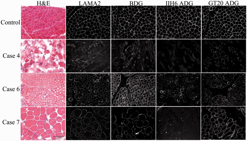
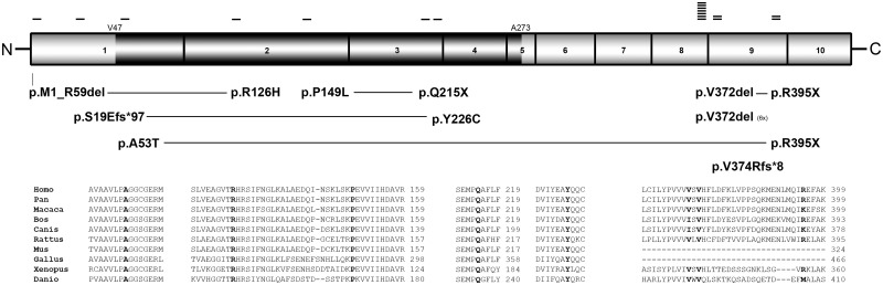
Dystroglycanopathies are a clinically and genetically diverse group of recessively inherited conditions ranging from the most severe of the congenital muscular dystrophies, Walker-Warburg syndrome, to mild forms of adult-onset limb-girdle muscular dystrophy. Their hallmark is a reduction in the functional glycosylation of α-dystroglycan, which can be detected in muscle biopsies. An important part of this glycosylation is a unique O-mannosylation, essential for the interaction of α-dystroglycan with extracellular matrix proteins such as laminin-α2. Mutations in eight genes coding for proteins in the glycosylation pathway are responsible for ∼50% of dystroglycanopathy cases. Despite multiple efforts using traditional positional cloning, the causative genes for unsolved dystroglycanopathy cases have escaped discovery for several years. In a recent collaborative study, we discovered that loss-of-function recessive mutations in a novel gene, called isoprenoid synthase domain containing (ISPD), are a relatively common cause of Walker-Warburg syndrome. In this article, we report the involvement of the ISPD gene in milder dystroglycanopathy phenotypes ranging from congenital muscular dystrophy to limb-girdle muscular dystrophy and identified allelic ISPD variants in nine cases belonging to seven families. In two ambulant cases, there was evidence of structural brain involvement, whereas in seven, the clinical manifestation was restricted to a dystrophic skeletal muscle phenotype. Although the function of ISPD in mammals is not yet known, mutations in this gene clearly lead to a reduction in the functional glycosylation of α-dystroglycan, which not only causes the severe Walker-Warburg syndrome but is also a common cause of the milder forms of dystroglycanopathy.


Figures

Figure 1
Figure 1
Clinical phenotype. Top: The clinical phenotype of ISPD-related muscular dystrophy in early childhood with pseudohypertrophy of lower limb muscles (A and B). Bottom: The disease at a later stage with marked pseudohypertrophy and ankle contractures (C, D and E). (A) Case 4 at 1.5 years, (B) Case 5 at 2.5 years, (C) Case 1 at 15.5 years, (D and E) Case 9 at 8 years.
Figure 2
Figure 2
Muscle MRI. (A) The T1-weighted muscle MRI of the thigh and calf of Case 1 at 19.5 years of age. (B) The T1-weighted muscle MRI of the thigh and calf of Case 9 at 20 years of age. There is sparing of sartorius and gracilis muscles (small arrows) in the thigh in both patients and sparing of the tibialis anterior muscle more visible in Case 1 (large arrow). The differential degree of fatty fibrous replacement is in concordance with the clinical severity with regards to ambulation, as Case 1 lost ambulation at 13 years, and Case 9 can still take a few steps with assistance only at age 23 years. AL = adductor longus; AM = adductor magnus; BF = biceps femoris; EDL = extensor digitorum longus; FDL = flexor digitorum longus; G = gracilis; GL = gastrocnemius lateralis; GM = gastrocnemius medialis; PG = peroneal group; RF = rectus femoris; S = sartorius; SM = semi-membranosus; SO = soleus; ST = semi-tendinosus; TA = tibialis anterior; TP = tibialis posterior; VL = vastus lateralis; VM = vastus medialis.
Figure 3
Figure 3
Brain MRI of Case 9 performed at 21 years of age, revealing brainstem hypoplasia, cerebellar hypoplasia and dilated fourth ventricle. (A) Sagittal T2-weighted image of the brain demonstrating brainstem hypoplasia, cerebellar hypoplasia and dilated fourth ventricle (arrow). (B) Axial T1-weighted image of the brain, revealing a normal appearance of the cerebral hemispheres. Interestingly, the temporalis muscles show some fibro-fatty changes (arrow). (C) Coronal T1-weighted image of the brain demonstrating dilatation of the fourth ventricle (arrow).
Figure 4
Figure 4
Immunohistochemistry of muscle biopsies from cases with ISPD-related muscular dystrophy. Unfixed frozen sections of quadriceps muscle biopsies from a control and three ISPD patients (Cases 4, 6, and 7) stained with haematoxylin and eosin (H & E) and immunolabelled with antibodies against laminin-α2 (LAMA2), β-dystroglycan (BDG), the glycosylated epitope of α-dystroglycan (IIH6 ADG) and the core protein of α-dystroglycan (GT20 ADG). Laminin-α2 immunostaining was reduced in all ISPD cases. Although expression of β-dystroglycan was similar to the control (see text), glycosylated α-dystroglycan immunolabelling was absent in Cases 4 and 7 and severely reduced in Case 6. Core α-DG (GT20ADG) was well preserved in all the cases.
Figure 5
Figure 5
ISPD protein and mutations. The domain structure of the ISPD protein is illustrated and the corresponding exons are shown. The darker region represents the active functionally conserved CDP-ME domain (V47–A273). Mutations found in a compound heterozygous state are connected with a horizontal line. The small-sized horizontal bars represent the number of observed alleles. Bottom: The conservation of the missense mutations (marked with bold letters) within the vertebrates [Homo sapiens NP_001094896.1; Pan troglodytes (XP_003318374.1) 99% amino acid identity; Macaca mulatta (XP_001105001.2) 94% amino acid identity; Bos taurus (XP_002686725.1) 81% amino acid identity; Canis lupus familiaris (XP_003431914.1) 84% amino acid identity; Rattus norvegicus (NP_001008387.1) 73% amino acid identity; Mus musculus (NP_848744.1) 74% amino acid identity; Gallus gallus (XP_418693.2) 66% amino acid identity; Xenopus tropicalis (NP_001016240.1) 60% amino acid identity; Danio rerio (NP_001071270.1) 47% amino acid identity].

Similar articles

Cited by

References

    1. Baur S, Marles-Wright J, Buckenmaier S, Lewis RJ, Vollmer W. Synthesis of CDP-activated ribitol for teichoic acid precursors in Streptococcus pneumoniae. J Bacteriol. 2009;191:1200–10. - PMC - PubMed
    1. Beltran-Valero de Bernabe D, Currier S, Steinbrecher A, Celli J, van Beusekom E, van der Zwaag B, et al. Mutations in the O-mannosyltransferase gene POMT1 give rise to the severe neuronal migration disorder Walker-Warburg syndrome. Am J Hum Genet. 2002;71:1033–43. - PMC - PubMed
    1. Bonnemann CG, Finkel RS. Sarcolemmal proteins and the spectrum of limb-girdle muscular dystrophies. Semin Pediatr Neurol. 2002;9:81–99. - PubMed
    1. Bourteel H, Vermersch P, Cuisset JM, Maurage CA, Laforet P, Richard P, et al. Clinical and mutational spectrum of limb-girdle muscular dystrophy type 2I in 11 French patients. Journal of neurology, neurosurgery, and psychiatry. 2009;80:1405–8. - PubMed
    1. Breton C, Snajdrova L, Jeanneau C, Koca J, Imberty A. Structures and mechanisms of glycosyltransferases. Glycobiology. 2006;16:29R–37R. - PubMed

Publication types

MeSH terms