. 2013 Jan:Chapter 22:Unit 22.1..
doi: 10.1002/0471142727.mb2201s101.

Overview of DNA microarrays: types, applications, and their future

Affiliations

Overview of DNA microarrays: types, applications, and their future

Roger Bumgarner. Curr Protoc Mol Biol. 2013 Jan.

Abstract

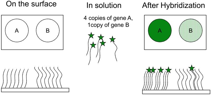
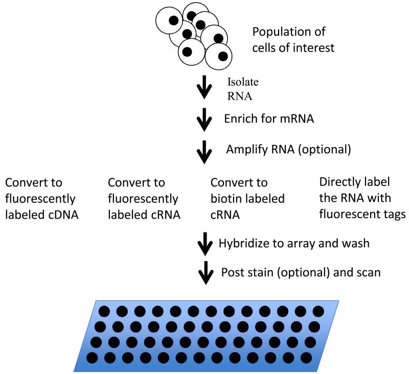
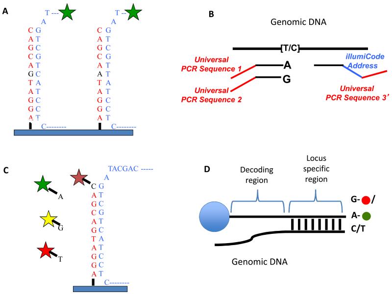
This unit provides an overview of DNA microarrays. Microarrays are a technology in which thousands of nucleic acids are bound to a surface and are used to measure the relative concentration of nucleic acid sequences in a mixture via hybridization and subsequent detection of the hybridization events. This overview first discusses the history of microarrays and the antecedent technologies that led to their development. This is followed by discussion of the methods of manufacture of microarrays and the most common biological applications. The unit ends with a brief description of the limitations of microarrays and discusses how microarrays are being rapidly replaced by DNA sequencing technologies.


Figures

Figure 1
Figure 1
Simplified view of a DNA array. The upper rectangles show two spots of DNA on a solid surface (sequences “A” and “B”) prior to and after hybridization. The lower rectangles show highly idealized side views of the same surfaces.
Figure 2
Figure 2
Three basic types of microarrays: (A) Spotted arrays on glass, (B) self assembled arrays and (C) in-situ synthesized arrays. A. With spotted arrays, a “pen” (or multiple pens) are dipped into solutions containing the DNA of interest and physically deposited on a 1“x 3” glass microscope slide. Typically the glass slide surface is coated with something to help retain the DNA such as polylysine {DeRisi, 1997 #28191}, a silane {Call, 2001 #28277} or a chemically reactive surface {Rogers, 1999 #28278} (to which chemically reactive oligos or PCR products would be added). B. Self assembled arrays can be created by applying a collection of beads containing a diverse set of oligos to a surface with pits the size of the beads. After the array is constructed a series of hybridizations determine which oligo is in what position on each unique array (Ferguson et al., 2000; Michael et al., 1998; Steemers et al., 2000; Walt, 2000) (Gunderson et al., 2004). C1 and C2. In-situ synthesized arrays can be produced by inkjet oligo synthesis methods (C1) or by photolithographic methods such as used by Affymetrix (C2).
Figure 3
Figure 3
Gene expression analysis via microarrays. RNA is isolated from the sample of interest and enriched for messenger RNA. In eucaryotes, poly-A tailed mRNA’s are typically enriched using affinity purification with oligo dT beads or columns. In procaryotes, unselected RNA is typically depleted for ribosomal sequences using bead or columns coated with sequences complementary to 16s. After message enriched RNA is in hand, it is optionally amplified and labeled by any one of a number of methods and the resulting labeled sample is hybridized to a microarray. The array is washed to remove unbound sample. If the sample was labeled with biotin, the array is post stained with fluorescently labeled streptavidin and washed again. The array is then scanned to measure fluorescence signal at each spot on the array.
Figure 4
Figure 4
SNP detections strategies for arrays. A) Allele discrimination by hybridization – Oligos that are complimentary to each allele are placed on the array and labeled genomic DNA is hybridized to the array. The variant position is placed in the center of the oligo (typically 25bp on Affymetrix arrays) as this position has the greatest affect on hybridization. Typically, multiple array positions are used for each allele to improve signal to noise. B) Illumina’s “Golden Gate Assay”- two allele specific oligos are each tailed with a different universal primer (1 and 2) and hybridized in solution to genomic DNA. A third oligo that is complementary to the same locus is tailed with a “barcode” sequence and a third universal primer (3). Polymerase is used to extend the allele specific primers across the genomic sequence and the extended products are ligated to the third oligo. PCR is performed using primers complimentary to universal sequences 1, 2 and 3. The PCR primers complimentary to the universal sequences 1 and 2 are labeled with a unique fluorophore. The barcode sequence on the third oligo allows the PCR product to be uniquely detected on an array containing oligos complimentary to the barcode sequence. The use of multiple barcodes (one for each locus of interest) allows the assay to be multiplexed to sample many loci. C) Arrayed primer extension (APEX) – In this assay, the array contains DNA oriented with the 5′ end attached to the array and the 3′ end stopping one nucleotide short of the SNP. Genomic DNA is fragmented and hybridized to the array and the oligo on the array is extended in single nucleotide dye terminator sequencing reaction. D) Illumina’s Infinium assay – This assay is similar to the APEX assay except that the oligo to be extended is on a bead and the single nucleotide that is added is labeled with a nucleotide specific hapten as opposed to a fluorophore. The haptens are then detected by staining with fluorescently labeled proteins that bind each hapten.

References

    1. Aaronson JS, Eckman B, Blevins RA, Borkowski JA, Myerson J, Imran S, Elliston KO. Toward the development of a gene index to the human genome: an assessment of the nature of high-throughput EST sequence data. Genome research. 1996;6:829–845. - PubMed
    1. Auffray C, Nageotte R, Chambraud B, Rougeon F. Mouse immunoglobulin genes: a bacterial plasmid containing the entire coding sequence for a pre-gamma 2a heavy chain. Nucleic acids research. 1980;8:1231–1241. - PMC - PubMed
    1. Auffray C, Rougeon F. Nucleotide sequence of a cloned cDNA corresponding to secreted mu chain of mouse immunoglobulin. Gene. 1980a;12:77–86. - PubMed
    1. Auffray C, Rougeon F. Purification of mouse immunoglobulin heavy-chain messenger RNAs from total myeloma tumor RNA. Eur J Biochem. 1980b;107:303–314. - PubMed
    1. Ball CA, Brazma A. MGED standards: work in progress. OMICS. 2006;10:138–144. - PubMed

Publication types

MeSH terms