Review
. 2013 Jan;43(1):15-27.
doi: 10.1007/s00247-012-2496-x. Epub 2013 Jan 4.

Diffusion tensor imaging of normal brain development

Affiliations
Review

Diffusion tensor imaging of normal brain development

Shoko Yoshida et al. Pediatr Radiol. 2013 Jan.

Abstract

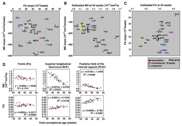
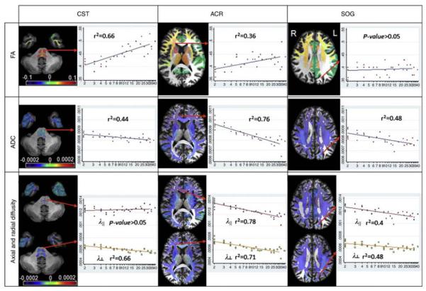
Diffusion tensor imaging (DTI) is an MRI technique that can measure the macroscopic structural organization in brain tissues. DTI has been shown to provide information complementary to relaxation-based MRI about the changes in the brain's microstructure. In the pediatric population, DTI enables quantitative observation of the maturation process of white matter structures. Its ability to delineate various brain structures during developmental stages makes it an effective tool with which to characterize both the normal and abnormal anatomy of the developing brain. This review will highlight the advantages, as well as the common technical pitfalls of pediatric DTI. In addition, image quantification strategies for various DTI-derived parameters and the normal brain developmental changes associated with these parameters are discussed.


Figures

Fig. 1
Fig. 1
The time course of normal maturation detected on T1 (upper row)/T2 (middle row) DTI (FA map, lower row). The maturation (predominantly the myelination pattern) has a profound impact on T1- and T2-weighted imaging. The extent of the contrast change is so large that the gray/white matter contrast inverts in the first 12 months. During this time period, FA remains more stable, especially in the deep brain regions, while the peripheral white matter has a noticeble FA increase
Fig. 2
Fig. 2
Multi-contrast, single-subject atlases for a neonate (with 112 parcellations) at 2 years old and 18 months old and in an adult (with 159 parcellations) (available at http://lbam.med.jhmi.edu/)]
Fig. 3
Fig. 3
Schematic pipeline of the image normalization process. The image on the upper left side is an initial image of children with periventricular leukomalacia, clinically diagnosed as spastic cerebral palsy. The orange arrows show “forward” transformation; the subject image was first linearly normalized (affine transformation), followed by nonlinear normalization (LDDMM). After this procedure, all subject images were transformed to a shape similar to that of the atlas (two images in lower left). For the “backward” transformation (green arrows), the brain parcellation map (BPM) was transformed to the original MRI using the same deformation fields used for the forward transformation. This allows the map to be superimposed onto the original images with parcellation into 159 structures. Please note the level of accuracy of the parcellation for this patient with severe injury
Fig. 4
Fig. 4
The relationship between the age-dependent mean diffusivity (MD) decreasing slope and the age-dependent fractional anisotropy (FA) increasing slope (a), the estimated MD at 40 post-conceptional weeks and the age-dependent MD decreasing slope (b), and the estimated FA at 40 post-conceptional weeks and the age-dependent FA increasing slope (c). The white matter structures were categorized based on the association fibers (black dots: SLF, SFO, ILF, IFO, and UNC), the commissural fibers (pink dots: CC and TAP), the limbic fibers (blue dots: CGC, CGH, Fx, and ST), and the projection fibers (green dots: ALIC, PLIC, RLIC, CP, PTR, SS, ACR, SCR, and PCR). (d) Linear regression analyses of MD and FA from three representive areas. Open squares indicates data from boys, and black circles indicate data from girls. The MD and FA of each structure show time-dependent changes with markedly different slopes and intercepts [71] (used by permission copyright). SLF superior longitudinal fasciculus, SFO superior fronto-occipital fasciculus, ILF inferior longitudinal fasciculus, IFO inferior fronto-occipital fasciculus, UNC uncinate fasciculus, CC corpus callosum, TAP tapetum, CGC cingulum cingular part, CGH cingulum hippocampal part, Fx fornix, ST stria terminalis, ALIC anterior limb of internal capsule, PLIC posterior limb of internal capsule, RLIC retrorenticular part of internal capsule, CP cerebral peduncle. PTR posterior thalamic radiation, SS sagittal stratum, ACR anterior corona radiata, SCR superior corona radiata, PCR posterior corona radiata
Fig. 5
Fig. 5
Actual fitting results for the fractional anisotropy (FA) (first row), apparent diffusion coefficient (ADC) (second row, in mm2/s), and axial and radial diffusivity (third row, in mm2/s), by age (in years, logarithmic scale), at representative locations. In the corticospinal tract (CST, first column), the FA increase can be explained by the radial diiffusivity decrease. In the anterior corona radiata (ACR, second column), the age-related changes in the axial diffusivity cause a weaker time-dependent FA change. In the white matter of the superior occipital gyrus (SOG, third column), the parallel decreases in both axial and radial diffusivity lead to no significant changes in FA. The orientation of the slices follows the radiologic convention (L left, R right) (printed with permission [72])
Fig. 6
Fig. 6
Map of slopes measured by the voxel- (a) and atlas-based analyses (b) for the volume, fractional anisotropy (FA), and diffusivity values. Note the overall agreement between the two methods. The orientation of the slices follows the radiologic convention (L left, R right. Th thalamus) (printed with permission [72])

References

    1. Barkovich AJ, Raybaud C. Pediatric neuroimaging. 5th edn. Lippincott Williams & Wilkins; Philadelphia: 2012.
    1. Keene MFL, Hewer EE. Some observations on myelination in the human nervous system. J Anat. 1931;6:1–13. - PMC - PubMed
    1. van der Knaap MS, Valk J. Magnetic resonance of myelination and myelin disorders. 3rd edn. Springer; Berlin: 2005.
    1. Yakovlev PI, Lecours AR. The myelogenetic cycles of regional maturation of the brain. In: Minkowski A, editor. Regional development of the brain in early life. Blackwell; Oxford: 1967.
    1. Ballesteros MC, Hansen PE, Solla K. MR imaging of the developing human brain. Part 2. Postnatal development. Radiographics. 1993;13:611–622. - PubMed

Publication types

LinkOut - more resources