. 2013 Mar 15;73(6):1733-41.
doi: 10.1158/0008-5472.CAN-12-2384. Epub 2013 Jan 3.

Evidence for a role of the PD-1:PD-L1 pathway in immune resistance of HPV-associated head and neck squamous cell carcinoma

Affiliations

Evidence for a role of the PD-1:PD-L1 pathway in immune resistance of HPV-associated head and neck squamous cell carcinoma

Sofia Lyford-Pike et al. Cancer Res. .

Abstract

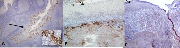
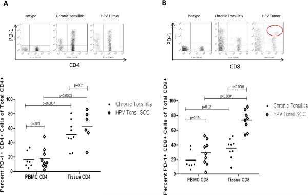
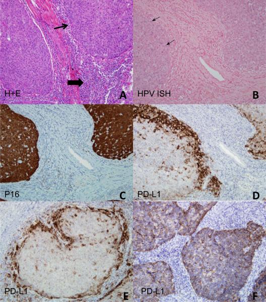
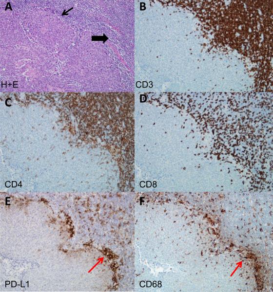
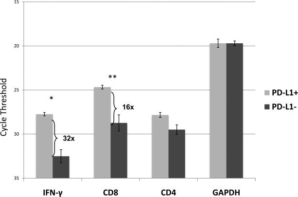
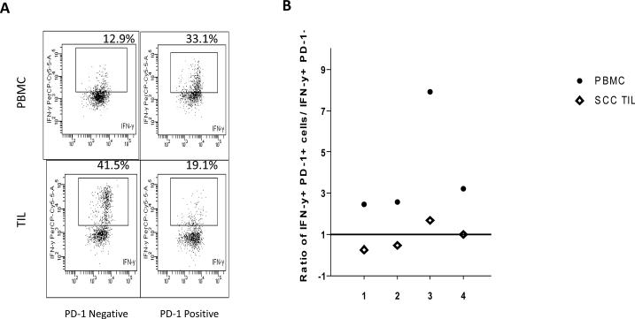
Human papillomavirus-associated head and neck squamous cell carcinomas (HPV-HNSCC) originate in the tonsils, the major lymphoid organ that orchestrates immunity to oral infections. Despite its location, the virus escapes immune elimination during malignant transformation and progression. Here, we provide evidence for the role of the PD-1:PD-L1 pathway in HPV-HNSCC immune resistance. We show membranous expression of PD-L1 in the tonsillar crypts, the site of initial HPV infection. In HPV-HNSCCs that are highly infiltrated with lymphocytes, PD-L1 expression on both tumor cells and CD68+ tumor-associated macrophages is geographically localized to sites of lymphocyte fronts, whereas the majority of CD8+ tumor-infiltrating lymphocytes express high levels of PD-1, the inhibitory PD-L1 receptor. Significant levels of mRNA for IFN-γ, a major cytokine inducer of PD-L1 expression, were found in HPV+ PD-L1(+) tumors. Our findings support the role of the PD-1:PD-L1 interaction in creating an "immune-privileged" site for initial viral infection and subsequent adaptive immune resistance once tumors are established and suggest a rationale for therapeutic blockade of this pathway in patients with HPV-HNSCC.


Figures

Figure 1
Figure 1. The reticulated epithelium of benign tonsil tissue expresses high levels of PD-L1
A. Chronically inflamed tonsil tissue demonstrates localized PD-L1 expression within the reticulated epithelium of tonsillar crypts (long arrow). Magnification, x40. The inset (magnification, x400) demonstrates cell surface staining of the crypt epithelial cells. In contrast, the surface epithelium of the tonsils was negative for PD-L1 expression (short arrows) (B, C). Magnification, x40.
Figure 2
Figure 2. High levels of PD-1 receptor on TILs from HPV-HNSCC
A. Representative flow cytometry of CD4+ T cell population expressing PD-1 in various tissues. Summary graph with mean frequency of CD4+PD-1(+) T cells in non-cancerous tonsils and HPV-HNSCC as compared to peripheral blood. B. Similar analysis performed for CD8+PD-1(+) T cell population. The circle on the representative flow data highlights a subpopulation of CD8+ TILs with high levels of PD-1 expression which was not observed in benign tonsils. (•) denotes chronic tonsillitis specimens and (◇) denotes HPV-HNSCC.
Figure 3
Figure 3. High levels of PD-L1 expression present in the tumor microenvironment of HPV-HNSCC
A. Hematoxylin and eosin stain of HPV-HNSCC demonstrates the proto-typic tumor nests (depicted by thin arrow) surrounded by a dense inflammatory stroma (depicted by thick arrow). Serial sections evaluated for B, HPV ISH (arrows mark areas of blue, intranuclear staining); C, p16 IHC; and D-F, PD-L1 IHC. Two patterns of PD-L1 staining were observed: peripheral tumoral staining (D, E) and diffuse intratumoral staining (F). Magnification, 400x
Figure 4
Figure 4. Co-localization of TILs with PD-L1 expression in HPV-HNSCC
A. Hematoxylin and eosin stain of HPV-HNSCC (thin arrow marks tumor nests and thick arrow marks inflammatory stroma). Serial sections evaluated for: B, CD3; C, CD4; D, CD8; E, PD-L1; and F, CD68 expression. Red arrows indicate a representative area with clusters of PD-L1 and CD68 expression in serial sections (E, F). Magnification, 400x.
Figure 5
Figure 5. Increased expression of IFN-γ and CD8 mRNA in PD-L1(+) vs. (-) tonsil cancers
IFN-γ, CD8, CD4, and GAPDH were evaluated by quantitative RT-PCR in both PD-L1(+) (n=3) and PD-L1(-) (n=6) tumors . Error bars represent standard error of the mean. Cycle threshold is on a log2 scale; lower numbers indicate greater expression. *p=0.003. **p=0.002. Results are representative of two separate experiments.
Figure 6
Figure 6. PD-1 expressing CD8+ TILs are functionally anergic relative to peripheral blood PD-1 expressing T cells
A. Representative flow cytometry comparing IFN-γ production by CD8+ T cells sorted by PD-1expression in PBMCs and TILs and stimulated with PMA/ionomycin. B. Summary graph of the ratio of IFN-γ production by PD-1(+) to PD-1(-) TILs and peripheral blood, in response to PMA/ionomycin. A value greater than 1 indicates increased IFN-γ production in PD-1(+) compared to PD-1(-) T cells, and a value less than 1 indicated a reduction. (•) denotes PBMC and (◇) denotes TILs.

References

    1. Gillison ML, Koch WM, Capone RB, Spafford M, Westra WH, Wu L, et al. Evidence for a causal association between human papillomavirus and a subset of head and neck cancers. J Natl Cancer Inst. 2000;92(9):709–20. - PubMed
    1. Chaturvedi Ak, Engels EA, Pfeiffer RM, Hernandez BY, Xiao W, Kim E, et al. Human papillomavirus and rising oropharyngeal cancer incidence in the United States. J Clin Oncol. 2011;1029(32):4294–301. - PMC - PubMed
    1. Begum S, Cao D, Gillison M, Zahurak M, Westra WH. Tissue distribution of human papillomavirus 16 DNA integration in patients with tonsillar carcinoma. Clin Cancer Res. 2005;11(16):694–9. - PubMed
    1. Singhi AD, Westra WH. Comparison of human papillomavirus in situ hybridization and p16 immunohistochemistry in the detection of human papillomavirus-associated head and neck acner based on a prospective clinical experience. Cancer. 2010;116(9):2166–73. - PubMed
    1. Gillison ML, D'Souza G, Westra W, Sugar E, Xiao W, Begum S, et al. Distinct risk factor profiles for human papillomavirus type 16-positive and human papilomavirus type 16-negative head and neck cancers. J Natl Cancer Inst. 2008;100(6):407–20. - PubMed

Publication types

MeSH terms