

Defining language networks from resting-state fMRI for surgical planning--a feasibility study
- PMID: 23288627
- PMCID: PMC3683367
- DOI: 10.1002/hbm.22231
Defining language networks from resting-state fMRI for surgical planning--a feasibility study
Abstract
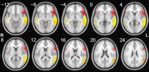
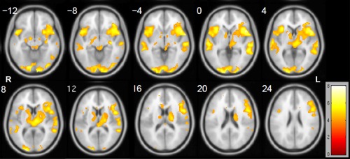
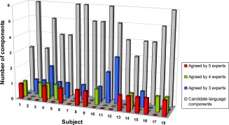
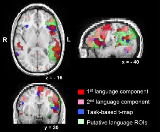
Presurgical language mapping for patients with lesions close to language areas is critical to neurosurgical decision-making for preservation of language function. As a clinical noninvasive imaging technique, functional MRI (fMRI) is used to identify language areas by measuring blood-oxygen-level dependent (BOLD) signal change while patients perform carefully timed language vs. control tasks. This task-based fMRI critically depends on task performance, excluding many patients who have difficulty performing language tasks due to neurologic deficits. On the basis of recent discovery of resting-state fMRI (rs-fMRI), we propose a "task-free" paradigm acquiring fMRI data when patients simply are at rest. This paradigm is less demanding for patients to perform and easier for technologists to administer. We investigated the feasibility of this approach in right-handed healthy control subjects. First, group independent component analysis (ICA) was applied on the training group (14 subjects) to identify group level language components based on expert rating results. Then, four empirically and structurally defined language network templates were assessed for their ability to identify language components from individuals' ICA output of the testing group (18 subjects) based on spatial similarity analysis. Results suggest that it is feasible to extract language activations from rs-fMRI at the individual subject level, and two empirically defined templates (that focuses on frontal language areas and that incorporates both frontal and temporal language areas) demonstrated the best performance. We propose a semi-automated language component identification procedure and discuss the practical concerns and suggestions for this approach to be used in clinical fMRI language mapping.
Keywords: functional connectivity; independent component analysis (ICA); language mapping; resting-state networks (RSNs); task-based fMRI; “task-free” paradigm.
Copyright © 2013 Wiley Periodicals, Inc.
Figures
References
-
- Auer DP ( (2008): Spontaneous low‐frequency blood oxygenation level‐dependent fluctuations and functional connectivity analysis of the ‘resting’ brain. Magn Reson Imaging 26:1055–1064. - PubMed
-
- Bandettini PA, Wong EC, Hinks RS, Tikofsky RS, Hyde JS ( (1992): Time course EPI of human brain function during task activation. Magn Reson Med 25:390–397. - PubMed
-
- Bartels A, Zeki S (2005): Brain dynamics during natural viewing conditions ‐A new guide for mapping connectivity in vivo. Neuroimage 24:339–349. - PubMed
-
- Bell AJ, Sejnowski TJ ( (1995): An information‐maximization approach to blind separation and blind deconvolution. Neural Comput 7:1129–1159. - PubMed
Publication types
MeSH terms
Grants and funding
LinkOut - more resources
Full Text Sources
Other Literature Sources
Research Materials
Miscellaneous
