. 2013 Apr;229(5):743-754.
doi: 10.1002/path.4158. Epub 2013 Mar 5.

Combining integrated genomics and functional genomics to dissect the biology of a cancer-associated, aberrant transcription factor, the ASPSCR1-TFE3 fusion oncoprotein

Affiliations

Combining integrated genomics and functional genomics to dissect the biology of a cancer-associated, aberrant transcription factor, the ASPSCR1-TFE3 fusion oncoprotein

Rachel Kobos et al. J Pathol. 2013 Apr.

Abstract

Oncogenic rearrangements of the TFE3 transcription factor gene are found in two distinct human cancers. These include ASPSCR1-TFE3 in all cases of alveolar soft part sarcoma (ASPS) and ASPSCR1-TFE3, PRCC-TFE3, SFPQ-TFE3 and others in a subset of paediatric and adult RCCs. Here we examined the functional properties of the ASPSCR1-TFE3 fusion oncoprotein, defined its target promoters on a genome-wide basis and performed a high-throughput RNA interference screen to identify which of its transcriptional targets contribute to cancer cell proliferation. We first confirmed that ASPSCR1-TFE3 has a predominantly nuclear localization and functions as a stronger transactivator than native TFE3. Genome-wide location analysis performed on the FU-UR-1 cell line, which expresses endogenous ASPSCR1-TFE3, identified 2193 genes bound by ASPSCR1-TFE3. Integration of these data with expression profiles of ASPS tumour samples and inducible cell lines expressing ASPSCR1-TFE3 defined a subset of 332 genes as putative up-regulated direct targets of ASPSCR1-TFE3, including MET (a previously known target gene) and 64 genes as down-regulated targets of ASPSCR1-TFE3. As validation of this approach to identify genuine ASPSCR1-TFE3 target genes, two up-regulated genes bound by ASPSCR1-TFE3, CYP17A1 and UPP1, were shown by multiple lines of evidence to be direct, endogenous targets of transactivation by ASPSCR1-TFE3. As the results indicated that ASPSCR1-TFE3 functions predominantly as a strong transcriptional activator, we hypothesized that a subset of its up-regulated direct targets mediate its oncogenic properties. We therefore chose 130 of these up-regulated direct target genes to study in high-throughput RNAi screens, using FU-UR-1 cells. In addition to MET, we provide evidence that 11 other ASPSCR1-TFE3 target genes contribute to the growth of ASPSCR1-TFE3-positive cells. Our data suggest new therapeutic possibilities for cancers driven by TFE3 fusions. More generally, this work establishes a combined integrated genomics/functional genomics strategy to dissect the biology of oncogenic, chimeric transcription factors.


Figures

Figure 1
Figure 1
(A) ASPSCR1, TFE3, ASPSCR1–TFE3 type1 and ASPSCR1–TFE3 type2 were cloned into a GFP vector and over-expressed in HeLa cells. Both types of ASPSCR1–TFE3 and native TFE3 showed predominantly nuclear localization, whereas native ASPSCR1 had a primarily cytoplasmic distribution. (B) To assess the activity of ASPSCR1–TFE3 as a TF, transactivation assays were performed using the µE3-luciferase reporter known to be bound by native TFE3. Both types of ASPSCR1–TFE3 were stronger activators of the μE3 reporter relative to TFE3. ASPSCR1–TFE3 type 2 was a stronger activator than ASPSCR1–TFE3 type 1 in 293 and Cos7 cells, but not in MCF-7 cells
Figure 2
Figure 2
Expression profiling data set on 139 sarcomas with chimeric TFs using Affymetrix U133A microarrays. Left panel shows unsupervised clustering of the expression data, demonstrating complete separation of the five sarcoma types in the dataset. The five sarcoma types and the corresponding gene fusions documented in all the cases are listed in the top right table. The bottom left table shows the analysis for differentially expressed genes in ASPS samples, all of which contained the ASPCR1–TFE3 fusion
Figure 3
Figure 3
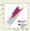
ChIP-on-chip results for ASPSCR1–TFE3-bound regions in the FU-UR-1 cell line. There were 2193 genes (11.5%) showing significant enrichment following IP for ASPSCR1–TFE3 using a TFE3 antibody in this cell line lacking native TFE3 expression
Figure 4
Figure 4
ChIP-on-chip results for MET, demonstrating the reproducibility of the results in the triplicate assays and providing basic validation of the overall experiment as the location of the four significant probes in ChIP-on-chip data coincides with the location of ASPSCR1–TFE3 binding (bracket in bottom panel), previously shown by simple ChIP analyses of the MET promoter region [15]
Figure 5
Figure 5
A. An analysis of over-represented 8 bp sequences in promoter regions bound by ASPSCR1–TFE3 rediscovered the CACGTG binding sequence which was previously described for TFE3, present here in four of the 10 top-scoring 8 bp motifs (boldface, red). In addition, it appeared to favour a 5'T and a 3'A. (B) Using MatrixREDUCE analysis, again the CACGTG consensus binding sequence was identified and favoured 5'T and 3'A. In this data visualization, the size and vertical order of the bases above the horizontal axis indicates the consensus and its strength at that position (see text for details)
Figure 6
Figure 6
There were a total of 332 genes (numbers in white) that were bound targets of ASPSCR1–TFE3 in the ChIP-on-chip analysis and up-regulated in either the ASPS sarcoma profiling signature (n = 93), the inducible 293 T cell signature (n = 174) or both (n = 65). We chose 130 of these 332 up-regulated ASPSCR1–TFE3 target genes to study in a high-throughput RNAi screen, based on biological plausibility and therapeutic potential. The final 12 genes emerging from this functional genomics screen are listed in Table 2. In addition, the 103 genes (65 + 38) that are in common between the ASPS sarcoma profiling signature and the inducible 293 T cell signature are listed in Table 1
Figure 7
Figure 7
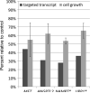
Individual validation of RNAi-mediated gene silencing of selected genes identified in the high-throughput primary screens. For each RNAi target, the percentage of transcript detected following knockdown is shown by the dark bar and the percentage of growth of FU-UR-1 cells following knockdown compared to mock-transfected control is shown by the light bar. *Cell viability assays following knockdowns of NAMPT and UPP1 were performed in the presence of 10 nM doxorubicin

Similar articles

Cited by

References

    1. Ladanyi M, Lui MY, Antonescu CR, et al. The der(17)t(X;17)(p11;q25) of human alveolar soft part sarcoma fuses the TFE3 transcription factor gene to ASPL, a novel gene at 17q25. Oncogene. 2001;20:48–57. - PubMed
    1. Argani P, Antonescu CR, Illei PB, et al. Primary renal neoplasms with the ASPL–TFE3 gene fusion of alveolar soft part sarcoma: a distinctive tumor entity previously included among renal cell carcinomas of children and adolescents. Am J Pathol. 2001;159:179–192. - PMC - PubMed
    1. Kayton ML, Meyers P, Wexler LH, et al. Clinical presentation, treatment, and outcome of alveolar soft part sarcoma in children, adolescents, and young adults. J Pediatr Surg. 2006;41:187–193. - PubMed
    1. Argani P, Olgac S, Tickoo SK, et al. Xp11 translocation renal cell carcinoma in adults: expanded clinical, pathologic, and genetic spectrum. Am J Surg Pathol. 2007;31:1149–1160. - PubMed
    1. Ishiguro M, Iwasaki H, Ohjimi Y, et al. Establishment and characterization of a renal cell carcinoma cell line (FU-UR-1) with the reciprocal ASPL–TFE3 fusion transcript. Oncol Rep. 2004;11:1169–1175. - PubMed

Publication types

MeSH terms

Substances