. 2013 Feb 15;288(7):5241-56.
doi: 10.1074/jbc.M112.404996. Epub 2013 Jan 3.

The regulated expression, intracellular trafficking, and membrane recycling of the P2Y-like receptor GPR17 in Oli-neu oligodendroglial cells

Affiliations

The regulated expression, intracellular trafficking, and membrane recycling of the P2Y-like receptor GPR17 in Oli-neu oligodendroglial cells

Alessandra Fratangeli et al. J Biol Chem. .

Abstract

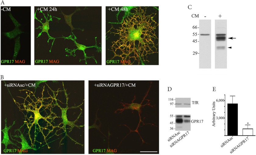
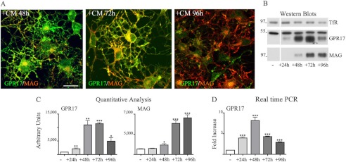
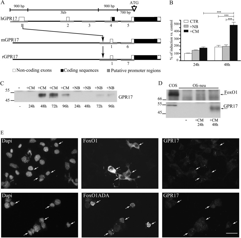
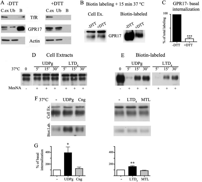
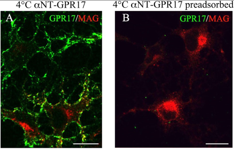
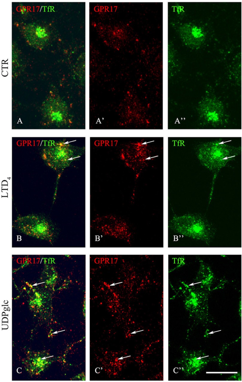
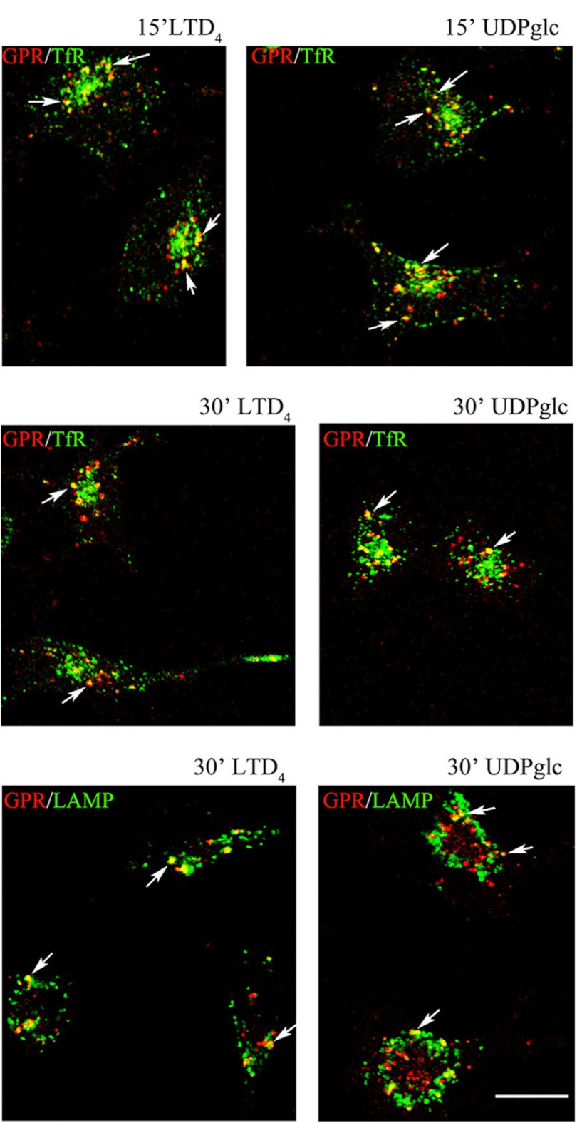
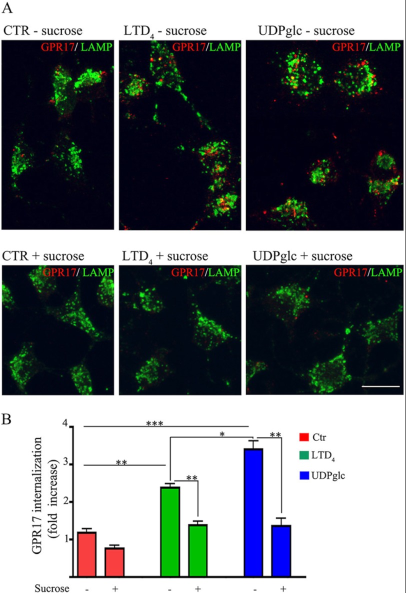
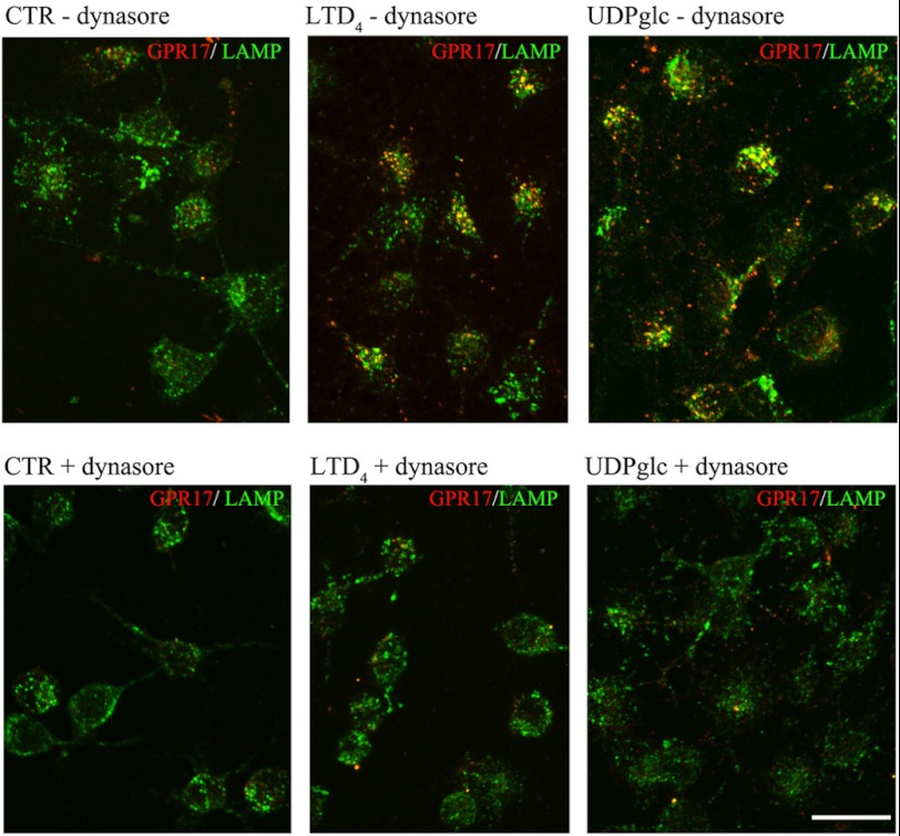
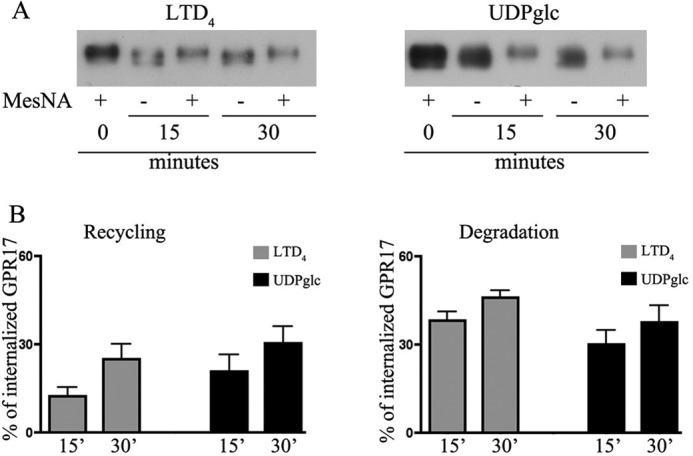
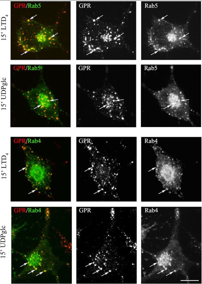
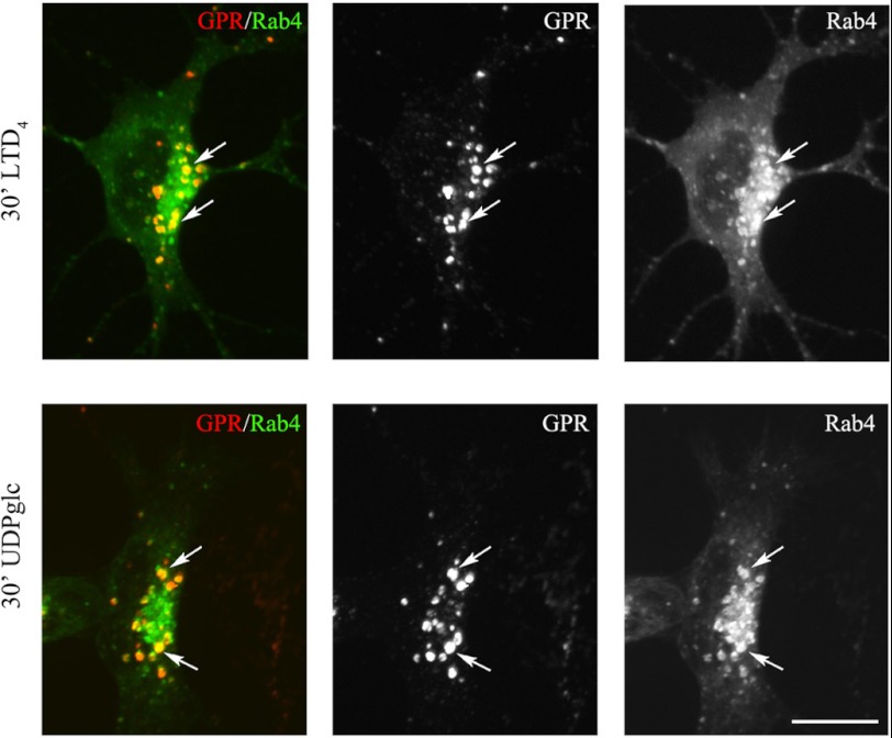
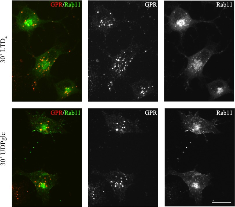
GPR17 is a G-protein-coupled receptor that is activated by two classes of molecules: uracil-nucleotides and cysteinyl-leukotrienes. GPR17 is required for initiating the differentiation of oligodendrocyte precursors but has to be down-regulated to allow cells to undergo terminal maturation. Although a great deal has been learned about GPR17 expression and signaling, no information is currently available about the trafficking of native receptors after the exposure of differentiating oligodendrocytes to endogenous agonists. Here, we demonstrate that neuron-conditioned medium induces the transcriptionally mediated, time-regulated expression of GPR17 in Oli-neu, an oligodendrocyte precursor cell line, making these cells suitable for studying the endocytic traffic of the native receptor. Agonist-induced internalization, intracellular trafficking, and membrane recycling of GPR17 were analyzed by biochemical and immunofluorescence assays using an ad hoc-developed antibody against the extracellular N-terminal of GPR17. Both UDP-glucose and LTD(4) increased GPR17 internalization, although with different efficiency. At early time points, internalized GPR17 co-localized with transferrin receptor, whereas at later times it partially co-localized with the lysosomal marker Lamp1, suggesting that a portion of GPR17 is targeted to lysosomes upon ligand binding. An analysis of receptor recycling and degradation demonstrated that a significant aliquot of GPR17 is recycled to the cell surface. Furthermore, internalized GPR17 displayed a co-localization with the marker of the "short loop" recycling endosomes, Rab4, while showing very minor co-localization with the "long loop" recycling marker, Rab11. Our results provide the first data on the agonist-induced trafficking of native GPR17 in oligodendroglial cells and may have implications for both physiological and pathological myelination.


Figures

FIGURE 1.
FIGURE 1.
GPR17 expression in Oli-neu cells. Oli-neu cells were cultured in Sato medium ([minus]CM) or conditioned medium (+CM, A) or transfected with scrambled siRNA (+siRNAsc) or a specific siRNA targeted against GPR17 (+siRNAGPR17) and then incubated in CM (+CM, B). At selected time points, the cells were fixed and double-immunolabeled with the anti-Ct-GPR17 antibody (∼1 μg/100 μl; green) and a monoclonal antibody against MAG (1:400, red) followed by fluorochrome-conjugated secondary antibodies. Images were recorded using a confocal microscope; merged images selected from four independent experiments are shown. Note the morphological changes and the increase in GPR17-labeling in Oli-neu cells maintained in medium supplemented with CM. Immunoreactivity for GPR17 was largely reduced upon transfection with a specific siRNA. Scale bar = 20 μm. In C, total membrane extracts (30 μg proteins) of Oli-neu cells cultured in Sato medium or CM (indicated as CM − or +) were analyzed by Western blotting using the anti-Ct-GPR17 antibody (1 μg/ml); the arrow and arrowhead indicate bands of 46–48 and 38 kDa, respectively, specifically immunodetected in differentiated cells. In D, total membrane extracts (30 μg) of Oli-neu cells cultured for 48 h in CM with scrambled siRNA (siRNAsc) or siRNAGPR17 were immunoblotted using antibodies against TfR (1:1000 dilution) or GPR17. Note the specific reduction of the 38 and 46–48 kDa bands after GPR17 knockdown, whereas Tfr and the 57-kDa band are unchanged. E, quantitative analysis of the intensity of the 46–48-kDa bands detected in Western blots of control and silenced cells; the data are expressed as arbitrary units and represent the mean values ±S.E. of three independent experiments (*, p < 0.05).
FIGURE 2.
FIGURE 2.
Synthesis and biochemical characterization of GPR17. A, immunoprecipitates with anti-Ct-GPR17 (αGPR) or preimmune rabbit IgG (pIg) from aliquots (100 μg of protein) of cell extracts from labeled (Lb) or 5 h “chased” (Ch) samples were analyzed by Western blotting followed by phosphorimaging analysis. Square brackets = 46–48-kDa bands; arrow = 38-kDa band. B, GPR17 glycosylation is shown. Protein (100 μg) from Triton X-100 extracts of undifferentiated (lanes 1 and 3) or 72 h-differentiated (lanes 2 and 4–8) Oli-neu cells were immunoprecipitated (IP) with anti-GPR17 or preimmune rabbit IgG (pIg). Before Western blot analysis, the samples in lanes 5–8 were incubated with endoglycosidase F (EdF) or H (EdH) at 37 or 0 °C. Square bracket = 46–48-kDa band, mature GPR17; arrow = 38-kDa band, immature GPR17; arrowhead = 33 kDa band un-glycosylated GPR17.
FIGURE 3.
FIGURE 3.
Transient expression of GPR17 in Oli-neu cells incubated in neuron-conditioned medium. In A, Oli-neu cells were cultured in CM for 48 h, 72 h, or 96 h (+CM) and then fixed and double-immunolabeled with antibodies against GPR17 or MAG (A′–C′). Images were recorded using a confocal microscope, and superimposed images (merges) are shown. Selected images from four independent experiments are shown. Scale bar = 20 μm. In B, cell extracts from Oli-neu cells cultured either in Sato medium (−) or in medium supplemented with CM (+) for 24, 48, 72, or 96 h were analyzed by Western blotting using antibodies against TfR, GPR17, or MAG (1:2,000 dilution). C, quantitative analysis of the Western blots from three independent experiments is shown; the intensity of the bands as measured by the ImageJ program is given as arbitrary units (*, p < 0.05; **, p < 0.01; ***, p < 0.0001). D, shown is real time PCR analysis of GPR17 gene expression in Oli-neu cells cultured in Sato medium (−) or exposed to CM (+) for the selected time points. GPR17 mRNA levels are relative to the housekeeping gene GAPDH and are expressed as -fold increase with respect to the mRNA levels (=1) detected in Oli-neu cells maintained in Sato medium. Data are from three independent experiments and represent the mean ± S.E. Statistical analysis was performed using a two-tailed, non-paired Student's t test (*, p < 0.05; **, p < 0.01; ***, p < 0.0001).
FIGURE 4.
FIGURE 4.
Analysis of purinergic and CysLT receptors in Oli-neu cells. Reverse transcription-PCR amplification was performed using specific primers (see “Experimental Procedures”) and 1 μg of RNA extracted from Oli-neu cells cultured in Sato medium alone (−) or in medium supplemented with CD (+) for 24 or 48 h. RNA from mouse brain (Br) and mouse platelets (Pl) was used as positive controls as indicated under “Experimental Procedures.” GAPDH was used as an internal standard. The final products were separated on an agarose gel (5 μl of the reaction mixtures were loaded on each lane). The bands revealed by ethidium bromide staining corresponded to the predicted sizes of the amplified DNA fragments.
FIGURE 5.
FIGURE 5.
The GPR17 promoter is selectively activated by CM. A, shown is a schematic representation of the human (h), mouse (m), and rat (r) GPR17 gene. The region upstream to the coding sequence has been analyzed with the Genomatix suite. Small blocks (from 600 bp to 1 kb) have been identified as putative promoter regions. Five regions in human, two in mouse, and two in rat were found (in gray). Only the region 1 is highly conserved among the three species. The human sequence 1 (909 bp) has been cloned in a pGL4 vector for the reporter assay. B, results of dual reporter luciferase assays show the GPR17 promoter activity in Oli-neu cells after transfection with the reporter construct pGL4-h909 or the corresponding empty vector. This was followed by incubation in Sato medium (control), neurobasal medium plus B27 (+NB), or CM for 24–48 h. Data are from three independent experiments and represent the mean ± S.E. Statistical analysis was performed using a two-way analysis of variance (***, p < 0.0001). C, cell extracts (30 μg protein) from Oli-neu cells incubated with either medium alone (−) or CM or neurobasal medium plus B27 (+NB) for the selected time points were analyzed by Western blotting using antibodies against GPR17. D, cell extracts from COS-7 (30 μg) or Oli-neu cells incubated with either Sato medium alone (−) or supplemented with CM for the selected time points were analyzed by Western blotting using antibodies against FoxO1 or GPR17. E, Oli-neu cells transiently transfected with cDNAs encoding for GFP-FoxO1 (FoxO1) or FLAG-FoxO1ADA were cultured in Sato medium for 48, fixed, and then labeled with anti-Ct-GPR17 antibodies and anti-FLAG antibody followed by secondary antibodies. Cells were examined using an Axiovert 200M confocal system equipped with a spinning disc. Arrows indicate FoxO1-transfected cells. Note that wild type protein is accumulated mainly in cytoplasm, whereas the constitutively active form (FoxO1ADA) is accumulated in the nucleus. The images are representatives from three independent experiments. Bar = 20 μm.
FIGURE 6.
FIGURE 6.
Agonist-induced internalization of GPR17. Internalization of GPR17 was measured in differentiated Oli-neu cells after cell surface labeling at 4 °C with the thiol-cleavable sulfo-NHS-SS-biotin. A, after labeling, cells were washed without (−) or with (+) DTT to remove the biotin from surface, and cell extracts (250 μg) were incubated with streptavidin. The proteins bound to streptavidin beads (B) and 5% of the cell extracts used for streptavidin-bead incubation (C.Ex) and also 5% of the supernatants from the streptavidin incubation (unbound proteins (Ub)) were analyzed by Western blotting with antibodies against TfR, GPR17, and actin. B, after labeling, Oli-neu cells were incubated for 15 min at 37 °C to allow for receptor internalization and then incubated without (−) or with (+) DTT. The cell extracts were analyzed as described above. In C, quantitative analysis of Western blots revealed that 10.82 ± 6.63% of the total biotinylated GPR17 underwent internalization under basal conditions. Data are from three independent experiments (the mean ± S.E.). ***, p < 0.0001. In D and E, after labeling cells were extracted or incubated with either UDP-glucose (100 μm, UDPg) or LTD4 (50 nm). At selected time points, the cells were cooled at 4 °C, incubated with the membrane-impermeable reducing agent MesNA to remove biotin from the remaining cell surface receptors, and solubilized. Aliquots (250 μg) of cell extracts were incubated with streptavidin beads and proteins bound to streptavidin (E) and also 10 μg of total cell extracts (D) were analyzed by Western blotting with antibodies against GPR17. F, after biotin labeling, Oli-neu cells were incubated at 37 °C in the absence (−) or presence of UDP-glucose, UDP-glucose (UDPg) and cangrelor (10 μm, Cng), LTD4, or LTD4 and montelukast (1 μm, MTL). After 15 min, cells were cooled at 4 °C, treated with reducing agents, and solubilized. Cell extracts (200 μg) were incubated with streptavidin beads, and bound proteins were analyzed by Western blotting with anti-GPR17 antibodies. The image shows representative blots from three independent experiments. G, graphs represent the quantitative analysis of blots, and the data are expressed as the percentage of biotinylated GPR17 internalized in the absence of agonists (−) and represent the mean ± S.E. (*, p < 0.05; **, p < 0.01, two-tailed, non-paired Student's t test).
FIGURE 7.
FIGURE 7.
Labeling of cell surface-exposed GPR17 with an antibody raised against the N terminus of GPR17. Cells were cultured with CM for 48 h and then incubated for 45 min at 4 °C with the anti-Nt-GPR17 antibody without or with preincubation with the antigen. After washing, the cells were fixed and labeled with a monoclonal antibody against MAG followed by an incubation with anti-mouse IgG conjugated to Cy3 (MAG, red) and an anti-rabbit IgG conjugated to fluorescein to reveal the distribution of the anti-GPR17 antibody (GPR17, green). Note that anti-Nt-GPR17 labels the surface of cells that do not express or express small amounts of MAG. Immunostaining for GPR17 was completely abolished by preincubating the antiserum with the peptide used for rabbit immunization. The merged images are shown. Bars = 10 μm.
FIGURE 8.
FIGURE 8.
Endocytosis of surface-labeled GPR17. Differentiated Oli-neu cells that were surface-labeled with the affinity-purified anti-Nt-GPR17 antibody were incubated without (control (CTR)) or with agonists (50 nm LTD4 or 100 μm UDP-glucose (UDPglc)). After an 8-min incubation at 37 °C, cells were washed with glycine buffer to remove the antibodies bound to the remaining cell surface-exposed receptors, fixed, and immunostained with a monoclonal antibody against TfR followed by incubation with anti-mouse IgG conjugated to fluorescein (TfR) and anti-rabbit IgG conjugated to Cy3 (GPR17). Confocal microscopy images show immunostaining for GPR17 was accumulated in small structures dispersed within the cytoplasm. Arrows indicate vesicles labeled for GPR17 and TfR. Images are representative of four independent experiments. The merged images are shown in A, B, and C. Bars = 10 μm.
FIGURE 9.
FIGURE 9.
GPR17 is accumulated in TfR- or Lamp1-positive compartments after 15 and 30 min of exposure to agonists. Differentiated Oli-neu cells were labeled with anti-Nt-GPR17 antibodies and incubated for 15 and 30 min at 37 °C with agonists (LTD4 or UDP-glucose (UDPglc)). After glycine washing, cells were fixed and labeled with monoclonal antibodies against TfR or Lamp1 followed by incubation with anti-rabbit IgG conjugated to Cy3 (GPR, red) and anti-mouse or anti-rat IgG conjugated to fluorescein (TfR or LAMP, green). Merged images represent individual confocal sections (0.5 μm) and are representative of four independent experiments. Note a subset of cytoplasmic structures co-labeled (yellow) for GPR17 and TfR or Lamp1. Bar = 10 μm.
FIGURE 10.
FIGURE 10.
GPR17 is internalized via clathrin-dependent endocytosis. In A, differentiated Oli-neu cells were surface-labeled with anti-Nt-GPR17 antibodies and then incubated for 12 min at 37 °C without (control (CTR)) or with agonists (LTD4 or UDP-glucose (UDPglc)) in Sato medium minus (−) or supplemented (+) with 450 mm sucrose. After glycine washing, cells were fixed and labeled with monoclonal antibodies against Lamp1 followed by incubation with anti-rat IgG conjugated to fluorescein (LAMP) and anti-rabbit IgG conjugated to Cy3 (GPR17). Confocal microscopy images show no accumulation of GPR17 immunostaining in the cells incubated in hypertonic sucrose. Images are representative of three independent experiments. The merged images are shown. Bar = 10 μm. B, to quantify the effects of clathrin lattice inhibition on the endocytosis of GPR17, confocal images were collected from Oli-neu cells incubated with or without hypertonic sucrose (+), and the pixel intensity was determined by ImageJ software. The graph represents the increase of GPR17 immunoreactivity in the cells over background (background = the immunofluorescence detected in the cells after labeling at 4 °C followed by acid stripping). The values are the mean (± S.E.) of three independent experiments. *, p > 0.05; **, p < 0.01; ***, p < 0.0001 (two-tailed, non-paired Student's t test).
FIGURE 11.
FIGURE 11.
Dynamin inhibition affects GPR17 endocytosis. Oli-neu cells were labeled with anti-Nt-GPR17 antibodies, pretreated at 4 °C with or without the dynamin inhibitor dynasore, and then incubated at 37 °C for 12 min in Sato medium (CTR) or medium supplemented with dynasore and UDP-glucose or LTD4. After fixation, cells were double-immunolabeled with monoclonal antibodies against Lamp1 followed by incubation with anti-rat IgG conjugated to fluorescein (LAMP, green) and anti-rabbit IgG conjugated to Cy3 (GPR17, red). Note the large reduction of GPR17 internalization in Oli-neu incubated with the inhibitor of dynamin (Bar = 20 μm).
FIGURE 12.
FIGURE 12.
Recycling and degradation of GPR17 after agonist-induced endocytosis. Oli-neu cells after cell surface labeling with sulfo-NHS-SS-biotin at 4 °C were incubated at 37 °C with UDP-glucose (100 μm, UDPglc) or LTD4 (50 nm). After 15 min, cells were cooled at 4 °C, incubated with MesNA to remove biotin from the remaining cell surface receptors, and solubilized (time 0) or further incubated in normal medium. At selected times, cells were either solubilized (−) or re-incubated with MesNA to remove biotinylated receptors re-exposed to the cell surface. In A aliquots (200 μg) of cell extracts were incubated with streptavidin beads, and the proteins bound to streptavidin were analyzed by Western blotting with antibodies against GPR17. In B, graphs represent the quantitative analysis of GPR17 recycling and degradation, which is reported as a percentage of the amount of receptor internalized after 15 min at 37 °C with LTD4 or UDP-glucose; the values are the mean of four independent experiments ±S.E.
FIGURE 13.
FIGURE 13.
At early time points endocytosed GPR17 accumulates in Rab5- or Rab4-positive vesicles. Oli-neu cells transiently transfected with cDNA encoding for GFP-Rab5a (Rab5) or GFP-Rab4a (Rab4) were labeled with anti-Nt-GPR17 antibodies and then stimulated with LTD4 or UDP-glucose (UDPglc) for 15 min. After glycine washing, the cells were fixed, and the endocytosed GPR17 was visualized by labeling with Cy3-conjugated antibodies (red). The cells were then examined using an Axiovert 200M confocal system equipped with a spinning disc. Note the co-localization of Rab proteins with GPR17 (arrows) in small vesicles scattered throughout the cytoplasm. The images are representatives from three independent experiments. Bar = 5 μm.
FIGURE 14.
FIGURE 14.
At later time points endocytosed GPR17 accumulates in Rab4-positive structures distributed in the perinuclear region. Oli-neu cells transiently transfected with cDNA encoding for GFP-Rab4a (Rab4) were labeled with anti-Nt-GPR17 antibodies and then stimulated with LTD4 or UDP-glucose (UDPglc) for 30 min. After glycine washing, cells were fixed, and the endocytosed GPR17 was visualized by labeling with Cy3-conjugated antibodies (GPR). The cells were examined using an Axiovert 200M confocal system equipped with a spinning disc. Note the co-localization of GPR17 with Rab4 (arrows) in dot-like structures distributed in the perinuclear region. The images are representatives from three independent experiments. Bar = 5 μm.
FIGURE 15.
FIGURE 15.
GPR17 exhibited a very minor degree of localization in Rab11-positive compartments. Oli-neu cells transiently transfected with cDNA encoding for GFP-Rab11a (Rab11) were labeled with anti-Nt-GPR17 antibodies and then stimulated with LTD4 or UDP-glucose (UDPglc) for 30 min. After glycine washing, the cells were fixed, and the endocytosed GPR17 was visualized by labeling with Cy3-conjugated antibodies (red, GPR). The cells were examined using an Axiovert 200M confocal system equipped with a spinning disc. The merged and single channel images shown are representatives from three independent experiments. Bar = 5 μm.

Similar articles

Cited by

References

    1. Ciana P., Fumagalli M., Trincavelli M. L., Verderio C., Rosa P., Lecca D., Ferrario S., Parravicini C., Capra V., Gelosa P., Guerrini U., Belcredito S., Cimino M., Sironi L., Tremoli E., Rovati G. E., Martini C., Abbracchio M. P. (2006) The orphan receptor GPR17 identified as a new dual uracil nucleotides/cysteinyl-leukotrienes receptor. EMBO J. 25, 4615–4627 - PMC - PubMed
    1. Lecca D., Trincavelli M. L., Gelosa P., Sironi L., Ciana P., Fumagalli M., Villa G., Verderio C., Grumelli C., Guerrini U., Tremoli E., Rosa P., Cuboni S., Martini C., Buffo A., Cimino M., Abbracchio M. P. (2008) The recently identified P2Y-like receptor GPR17 is a sensor of brain damage and a new target for brain repair. PLoS One 3, e3579. - PMC - PubMed
    1. Parravicini C., Ranghino G., Abbracchio M. P., Fantucci P. (2008) GPR17. Molecular modeling and dynamics studies of the 3-D structure and purinergic ligand binding features in comparison with P2Y receptors. BMC Bioinformatics 9, 263–282 - PMC - PubMed
    1. Pugliese A. M., Trincavelli M. L., Lecca D., Coppi E., Fumagalli M., Ferrario S., Failli P., Daniele S., Martini C., Pedata F., Abbracchio M. P. (2009) Functional characterization of two isoforms of the P2Y-like receptor GPR17. [35S]GTPγS binding and electrophysiological studies in 1321N1 cells. Am. J. Physiol. Cell. Physiol. 297, C1028–C1040 - PubMed
    1. Daniele S., Lecca D., Trincavelli M. L., Ciampi O., Abbracchio M. P., Martini C. (2010) Regulation of PC12 cell survival and differentiation by the new P2Y-like receptor GPR17. Cell. Signal. 22, 697–706 - PubMed

Publication types