. 2013 Jan 15;110(3):1101-6.
doi: 10.1073/pnas.1119416110. Epub 2013 Jan 3.

Reversal of end-stage retinal degeneration and restoration of visual function by photoreceptor transplantation

Affiliations

Reversal of end-stage retinal degeneration and restoration of visual function by photoreceptor transplantation

Mandeep S Singh et al. Proc Natl Acad Sci U S A. .

Abstract

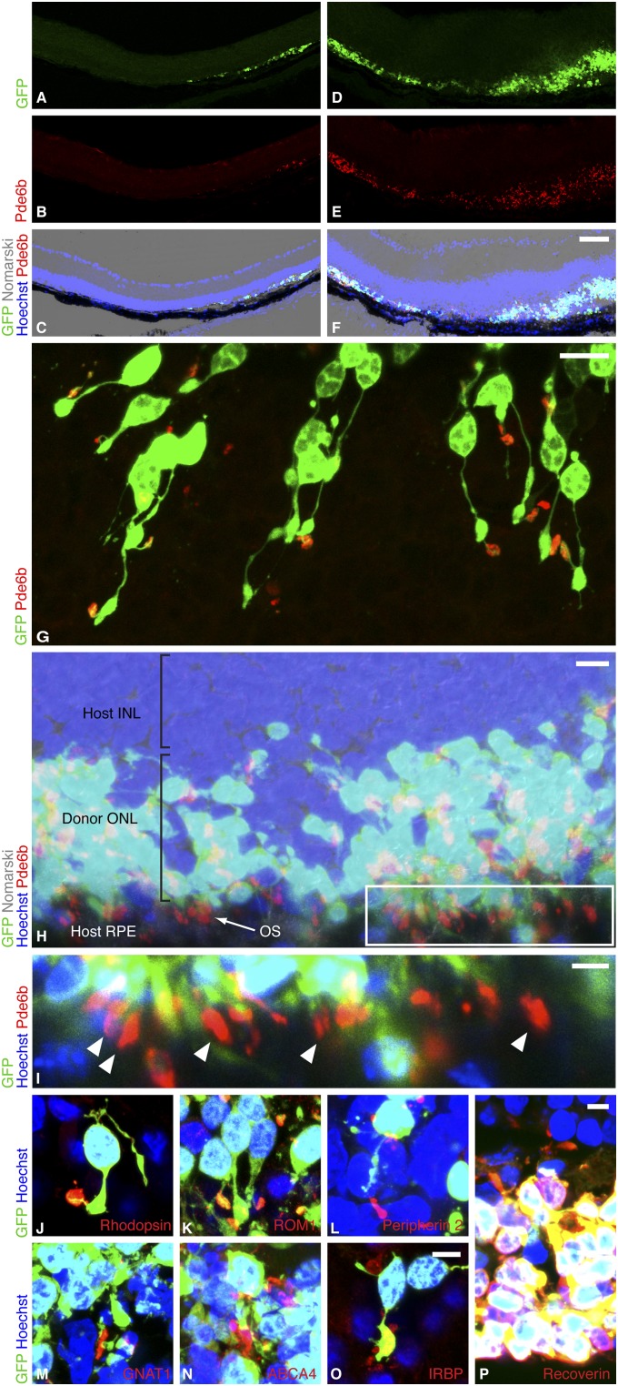
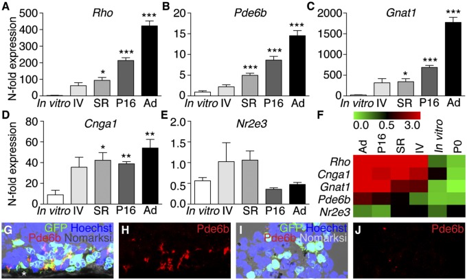
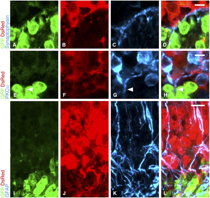
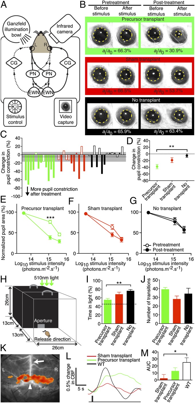
One strategy to restore vision in retinitis pigmentosa and age-related macular degeneration is cell replacement. Typically, patients lose vision when the outer retinal photoreceptor layer is lost, and so the therapeutic goal would be to restore vision at this stage of disease. It is not currently known if a degenerate retina lacking the outer nuclear layer of photoreceptor cells would allow the survival, maturation, and reconnection of replacement photoreceptors, as prior studies used hosts with a preexisting outer nuclear layer at the time of treatment. Here, using a murine model of severe human retinitis pigmentosa at a stage when no host rod cells remain, we show that transplanted rod precursors can reform an anatomically distinct and appropriately polarized outer nuclear layer. A trilaminar organization was returned to rd1 hosts that had only two retinal layers before treatment. The newly introduced precursors were able to resume their developmental program in the degenerate host niche to become mature rods with light-sensitive outer segments, reconnecting with host neurons downstream. Visual function, assayed in the same animals before and after transplantation, was restored in animals with zero rod function at baseline. These observations suggest that a cell therapy approach may reconstitute a light-sensitive cell layer de novo and hence repair a structurally damaged visual circuit. Rather than placing discrete photoreceptors among preexisting host outer retinal cells, total photoreceptor layer reconstruction may provide a clinically relevant model to investigate cell-based strategies for retinal repair.


Conflict of interest statement

The authors declare no conflict of interest.

Figures

Fig. 1.
Fig. 1.
Rod precursors transplanted into the degenerate host regenerate a new photoreceptor layer. (A–F) Two regions of rd1 host retina after subretinal transplantation of P3 Tg(Nrl-L-EGFP) rod precursors. By 2 wk, the retina had reattached and a new donor-derived ONL, marked by GFP, was formed. AC show an area where one to three ONL rows were formed, and DF show up to 10 rows. An internal nontransplant control region lacking an ONL is shown in the left half of AC. (Scale bar, 75 µm.) (G) As a sign of maturation, donor-derived GFP-positive rods formed slender processes extending from the cell body and expressed phosphodiesterase β6 (Pde6b) sequestered in discrete OS. (Scale bar, 10 µm.) (H) In many regions, Pde6b-positive OS were appropriately polarized with respect to host RPE, which is the optimal configuration for rod OS maintenance. (Scale bar, 10 µm.) Boxed region of H enlarged in I shows OS (arrowheads) aligned in rows against host RPE. (Scale bar, 5 µm.) (JN) As further evidence of maturation, rhodopsin, rod outer segment membrane protein 1 (ROM1), peripherin 2, guanine nucleotide-binding protein G(t) subunit α1 (GNAT1), and ATP-binding cassette subfamily A member 4 (ABCA4) were localized in outer segments. (O) Donor cells expressed interphotoreceptor retinoid-binding protein (IRBP), a secreted protein critical for ONL maintenance, and (P) were positive for the photoreceptor marker recoverin. (Scale bar, 5 µm for JP.)
Fig. 2.
Fig. 2.
Developmental cues support the maturation of rod precursors in the degenerate subretinal niche. (A–D) Rod-specific gene expression, measured by qRT-PCR. P2–3 Tg(Nrl-L-EGFP) retinal cells were exposed to different conditions [transplanted intravitreally (IV), transplanted subretinally (SR), maintained in vitro, or allowed to complete development in situ until P16]. Genes expressed highly in P16 and adult (Ad) Tg(Nrl-L-EGFP) eyes and vital for rod outer segment homeostasis were also highly expressed by precursors transplanted subretinally into degenerate rd1 hosts. Rho (F4,12.37 = 86.12, P = 6.5 × 10⁻⁹), Pde6b (F4,14.41 = 41.60, P = 9.3 × 10⁻⁸), Gnat1 (F4,12.41 = 87.89, P = 5.5 × 10⁻⁹), Cnga1 (F4,12.23 = 9.58, P = 0.001). *P < 0.05, **P < 0.01, ***P < 0.001 post hoc versus in vitro group, n = 5–8 per niche condition. Expression of these genes in P2–3 cells transplanted intravitreally showed a trend of being up-regulated compared with in vitro controls, but this did not reach statistical significance. Error bars, SEM. (E) Nr2e3 is a developmentally regulated rod transcription factor gene that peaks during early rod genesis around the time of transplantation and was used as an internal control. This was not up-regulated relative to in vitro controls (F4,29 = 2.47, P = 0.07), which confirmed that the increased gene expression was specific for late rod development. Data are presented as fold change in mRNA expression compared with expression in P0 retina (n = 7). (F) Heat map colors represent the expression of each gene individually in the different niches and are not comparable between genes. (G and H) Pde6b-positive OS in subretinally transplanted precursors. Asterisk, retinal pigment epithelium. (I and J) Poor OS formation after intravitreal transplantation.
Fig. 3.
Fig. 3.
Integration of transplanted cells with host inner retina. (AD) Synaptophysin (blue) expression was appropriately located between host INL marked by DsRed (red) expression and the donor-derived ONL marked by GFP (green). (EH) Host bipolar cells, marked with PKCα (blue), extended processes (arrowheads) into the graft. (Scale bars, 5 µm for AH.) (IL) Host Müller cells are identified by the coexpression of DsRed (red) and GFAP (blue). Müller cell processes extend from the host INL into the graft (green). (Scale bar, 10 µm.)
Fig. 4.
Fig. 4.
Rod precursor transplantation restores visual function to eyes with complete rod degeneration. (A) Retinal illumination results in signal transmission to both pretectal nuclei (PN) and Edinger–Westphal nuclei (EWN). Impulses are relayed via the ciliary ganglia (CG) and both pupils constrict. Solid and dashed lines show reflex arcs triggered by left (L) and right (R) eye stimulation, respectively. (B) Representative pupil images. (C) Overview of the change in pupil constriction from before to after treatment in all mice at 1.21 × 10¹⁵ photons⋅cm−2⋅s−1. Green, red, and black denote precursor, sham, and no transplant controls, respectively. Dots indicate values close to zero and dashed line indicates the mean change with 95% confidence interval (gray) of the non-transplanted controls. The PLR was measured in the same eye 3 d before and 2 wk after subretinal transplantation of 3 × 10⁵ cells. PLR was expressed as normalized pupil area as previously described (3). (D) Precursor transplantation improved pupil constriction, compared with no transplant controls (F2,34 = 7.264, P = 0.002, n = 11–14 each; **P = 0.001 post hoc). (E) Paired analysis before versus after treatment showed that at the median light stimulus intensity, the PLR improved significantly in precursor-treated animals (***t11 = 5.95, P < 0.0001, n = 12). (F and G) The PLR did not improve after sham transplantation (n = 11) or in untreated controls (n = 14) at any stimulus intensity. There were no significant differences between groups at baseline (P = 0.12). (H) Light-mediated behavior was measured under dim 510-nm illumination (150 nW⋅cm−2⋅s−1). (I) Precursor-transplanted mice spent less time in the lit compartment than rd1 controls (F2,20 = 7.608, P = 0.003, n = 7–8 each; **P = 0.003 post hoc). Dashed line indicates mean WT level. (J) Anxiety-related behavior (transitions between compartments) was similar across groups. (K) Cortical activation map in WT showing increased cortical blood flow (CBF) over L and R visual cortices; arrow, superior sagittal sinus; arrowhead, lambda. (L) Averaged L and R CBF time series from all mice after a 4-Hz (1-s) stimulus (black bar). (M) Precursor transplantation led to a higher CBF change than sham treatment compared with WT (F2,16 = 4.108, P = 0.04, n = 5–7 each; *P = 0.01). AUC, area under curve (0–7 s). Error bars, SEM.

References

    1. Zrenner E, et al. Subretinal electronic chips allow blind patients to read letters and combine them to words. Proc Biol Soc. 2011;278(1711):1489–1497. - PMC - PubMed
    1. Eiraku M, et al. Self-organizing optic-cup morphogenesis in three-dimensional culture. Nature. 2011;472(7341):51–56. - PubMed
    1. MacLaren RE, et al. Retinal repair by transplantation of photoreceptor precursors. Nature. 2006;444(7116):203–207. - PubMed
    1. Bowes C, et al. Retinal degeneration in the rd mouse is caused by a defect in the β subunit of rod cGMP-phosphodiesterase. Nature. 1990;347(6294):4. - PubMed
    1. Akimoto M, et al. Targeting of GFP to newborn rods by Nrl promoter and temporal expression profiling of flow-sorted photoreceptors. Proc Natl Acad Sci USA. 2006;103(10):3890–3895. - PMC - PubMed

Publication types