. 2013 Feb;154(2):738-48.
doi: 10.1210/en.2012-1790. Epub 2013 Jan 3.

Developmental analysis and influence of genetic background on the Lhx3 W227ter mouse model of combined pituitary hormone deficiency disease

Affiliations

Developmental analysis and influence of genetic background on the Lhx3 W227ter mouse model of combined pituitary hormone deficiency disease

Kelly L Prince et al. Endocrinology. 2013 Feb.

Abstract

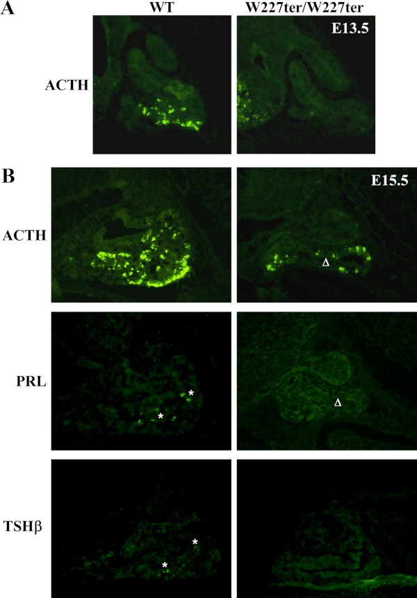
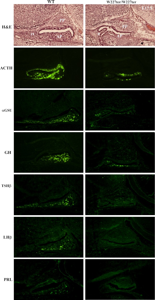
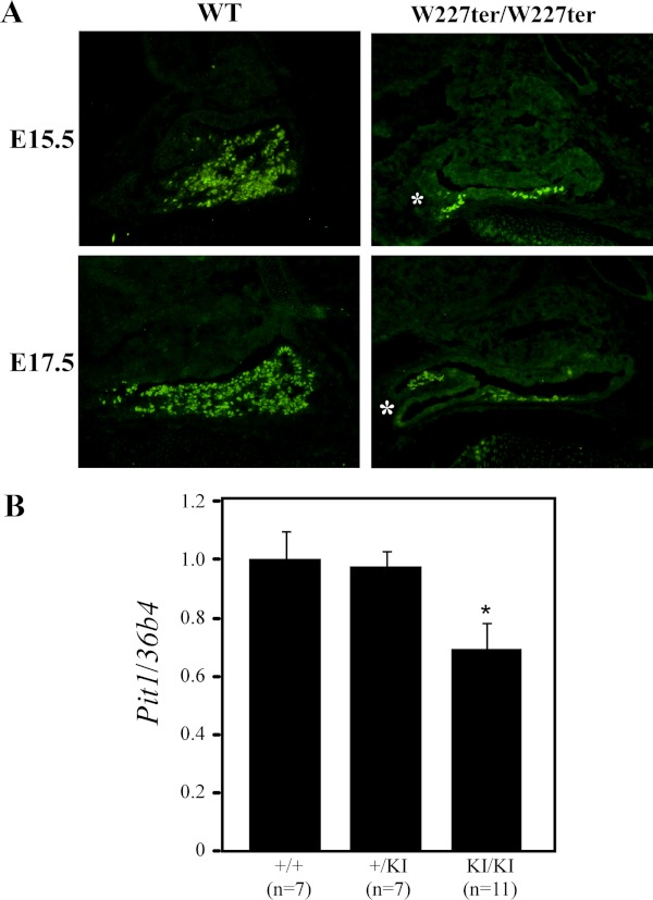
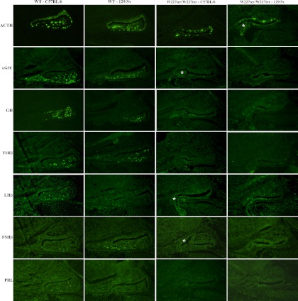
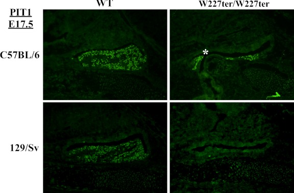
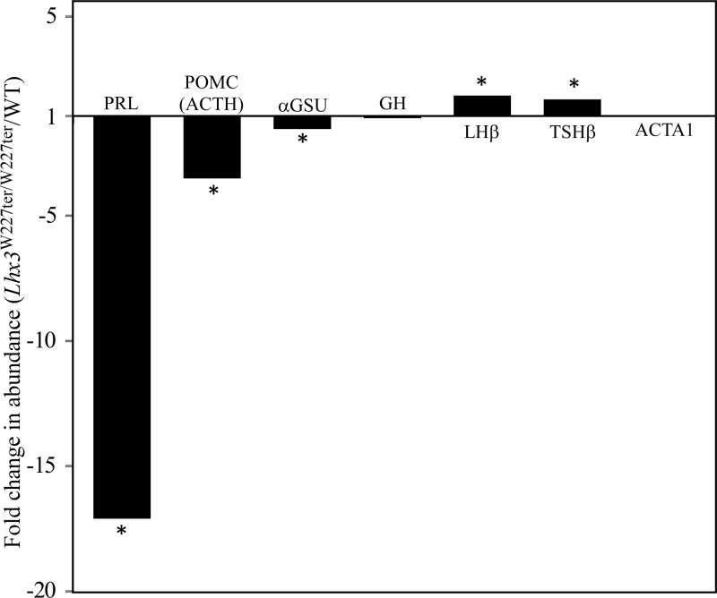
Combined pituitary hormone deficiency (CPHD) diseases result in severe outcomes for patients including short stature, developmental delays, and reproductive deficiencies. Little is known about their etiology, especially the developmental profiles and the influences of genetic background on disease progression. Animal models for CPHD provide valuable tools to investigate disease mechanisms and inform diagnostic and treatment protocols. Here we examined hormone production during pituitary development and the influence of genetic background on phenotypic severity in the Lhx3(W227ter/W227ter) mouse model. Lhx3(W227ter/W227ter) embryos have deficiencies of ACTH, α-glycoprotein subunit, GH, PRL, TSHβ, and LHβ during prenatal development. Furthermore, mutant mice have significant reduction in the critical pituitary transcriptional activator-1 (PIT1). Through breeding, the Lhx3(W227ter/W227ter) genotype was placed onto the 129/Sv and C57BL/6 backgrounds. Intriguingly, the genetic background significantly affected viability: whereas Lhx3(W227ter/W227ter) animals were found in the expected frequencies in C57BL/6, homozygous animals were not viable in the 129/Sv genetic environment. The hormone marker and PIT1 reductions observed in Lhx3(W227ter/W227ter) mice on a mixed background were also seen in the separate strains but in some cases were more severe in 129/Sv. To further characterize the molecular changes in diseased mice, we conducted a quantitative proteomic analysis of pituitary proteins. This showed significantly lower levels of PRL, pro-opiomelanocortin (ACTH), and α-glycoprotein subunit proteins in Lhx3(W227ter/W227ter) mice. Together, these data show that hormone deficiency disease is apparent in early prenatal stages in this CPHD model system. Furthermore, as is noted in human disease, genetic background significantly impacts the phenotypic outcome of these monogenic endocrine diseases.


Figures

Figure 1.
Figure 1.
Delayed onset of AP hormone expression in Lhx3W227ter/W227ter mice during development. ACTH, PRL, and TSHβ hormones were labeled using fluorescent IHC in pituitary sections of homozygote Lhx3W227ter/W227ter and WT mice at E15.5 (n = 5 per group). ACTH was also labeled in E13.5 sections (n = 5 per group). A and B, No specific ACTH expression was observed in Lhx3W227ter/W227ter animals at E13.5, and ACTH expression was decreased at E15.5. B, Low levels of PRL and TSHβ (asterisks) were detectable at E15.5, but no specific staining was seen in Lhx3W227ter/W227ter mutants. Lhx3W227ter/W227ter anterior pituitary lobes often were hypoplastic compared with WT controls (eg, Δ).
Figure 2.
Figure 2.
The expression of AP hormones is reduced or absent in embryonic Lhx3W227ter/W227ter mice. ACTH, αGSU, GH, TSHβ, LHβ, and PRL pituitary hormone markers were labeled using IHC in E17.5 pituitary sections of Lhx3W227ter/W227ter homozygote and WT mice. Decreased expression of ACTH, αGSU, and GH was observed for Lhx3W227ter/W227ter animals, and no labeling was detected for TSHβ, LHβ, and PRL (n = 5 per group). Hematoxylin and eosin (H&E) staining (top panels) revealed that Lhx3W227ter/W227ter APs often were hypoplastic with structural abnormalities such as bifurcations (asterisk). PP indicates the posterior lobe; rt, rostral tip.
Figure 3.
Figure 3.
Expression of the PIT1 pituitary transcription factor is reduced in Lhx3W227ter/W227ter animals. A, PIT1 protein was labeled using IHC in pituitary sections of Lhx3W227ter/W227ter homozygote and WT mice at E15.5 and E17.5. Lhx3W227ter/W227ter animals had decreased amounts of nuclear staining in PIT1-positive cells (n = 5 per group). Examples of a hypoplastic, bifurcated AP lobe also are noted (asterisks). B, Measurement of Pit1/Pou1f1 transcript levels in pituitary glands of 12-week-old adult mice using real-time quantitative PCR revealed a significant decrease in levels in the Lhx3W227ter/W227ter homozygote knock-in (KI/KI) mice. The asterisk indicates significance when compared with WT controls with P < .05. Data were normalized by determining the relative abundance of 36B4 (acidic ribosomal phosphoprotein P0 [Rplp0 gene]) mRNA. Examples of hypoplastic, bifurcated anterior pituitary lobes are noted (*, asterisks).
Figure 4.
Figure 4.
Genetic background impacts the expression of AP hormones in the Lhx3W227ter/W227ter mouse. Pituitary hormone markers were analyzed by IHC in animals on C57BL/6 and 129/Sv backgrounds. AP hormone expression in Lhx3W227ter/W227ter homozygote knock-in and WT mice at E17.5 on a 98.4% C57BL/6 or 129/Sv genetic background. Lhx3W227ter/W227ter animals had decreased expression of ACTH and αGSU and no expression of PRL, FSHβ, TSHβ, or LHβ in both strains. Neither Lhx3W227ter/W227ter nor WT animals on a 129/Sv background had any PRL labeling. Lhx3W227ter/W227ter animals on a 129/Sv background had no GH expression, whereas those on a C57BL/6 background had decreased expression of GH compared with WT. Examples of hypoplastic, bifurcated AP lobes are noted (asterisks).
Figure 5.
Figure 5.
Reduced expression of PIT1 protein in Lhx3W227ter/W227ter animals on C57BL/6 and 129/Sv backgrounds. PIT1 protein was detected using IHC in pituitary sections of Lhx3W227ter/W227ter homozygote and WT mice at E17.5 on a 98.4% C57BL/6 or 129/Sv background. Decreased expression was seen in Lhx3W227ter/W227ter animals on a C57BL/6 background, and no expression was seen in Lhx3W227ter/W227ter mice on a 129/Sv background. An example of a hypoplastic, bifurcated AP lobe is labeled (asterisk).
Figure 6.
Figure 6.
Proteomic analysis of pituitary hormone markers in Lhx3W227ter/W227ter animals. Pituitaries were harvested from adult female Lhx3W227ter/W227ter or WT animals, and protein extracts were analyzed by label-free quantitative liquid chromatography tandem mass spectroscopy (see Materials and Methods). Fold changes in relative abundances of PRL, POMC (representing ACTH), αGSU (from the mouse Cga gene), LHβ, TSHβ, GH, and ACTA1 (α-actin, as a comparator negative control) proteins/peptides were calculated (Lhx3W227ter/W227ter homozygote/WT). PRL, POMC, and αGSU were significantly lower in Lhx3W227ter/W227ter animals at −17.1-fold, −3.5-fold, and −1.6-fold respectively. (Note that in proteomic method development, the PRL result depicted here was shown as an isolated example for the technique.) (32). GH showed no significant change. LHβ and TSHβ were increased in Lhx3W227ter/W227ter animals at 1.8-fold and 1.7-fold, respectively. The control ACTA1 showed no significant change. Asterisks indicate significance at P < .00876 (q < 0.05) (n = 5 per group).

Similar articles

Cited by

References

    1. Kelberman D, Rizzoti K, Lovell-Badge R, Robinson IC, Dattani MT. Genetic regulation of pituitary gland development in human and mouse. Endocr Rev. 2009;30:790–829 - PMC - PubMed
    1. Davis SW, Castinetti F, Carvalho LR, et al. Molecular mechanisms of pituitary organogenesis: In search of novel regulatory genes. Mol Cell Endocrinol. 2010;323:4–19 - PMC - PubMed
    1. Mollard P, Hodson DJ, Lafont C, Rizzoti K, Drouin J. A tridimensional view of pituitary development and function. Trends Endocrinol Metab. 2012;23:261–269 - PubMed
    1. Prince KL, Walvoord EC, Rhodes SJ. The role of homeodomain transcription factors in heritable pituitary disease. Nat Rev Endocrinol. 2011;7:727–737 - PubMed
    1. Sharma K, Sheng HZ, Lettieri K, et al. LIM homeodomain factors Lhx3 and Lhx4 assign subtype identities for motor neurons. Cell. 1998;95:817–828 - PubMed

Publication types