. 2013 Mar;54(3):581-591.
doi: 10.1194/jlr.M027060. Epub 2013 Jan 2.

Cell wounding activates phospholipase D in primary mouse keratinocytes

Affiliations

Cell wounding activates phospholipase D in primary mouse keratinocytes

Senthil N Arun et al. J Lipid Res. 2013 Mar.

Abstract

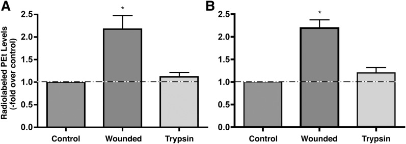
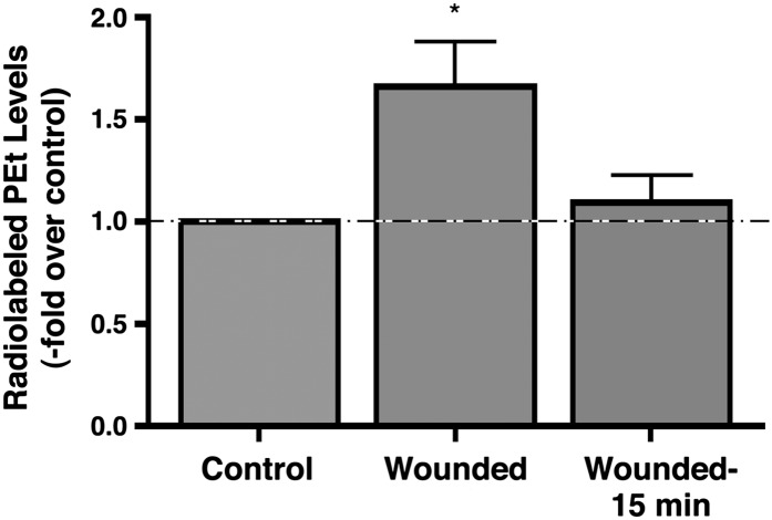
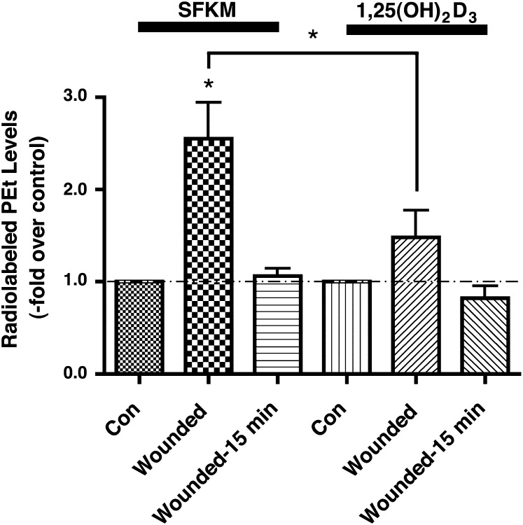
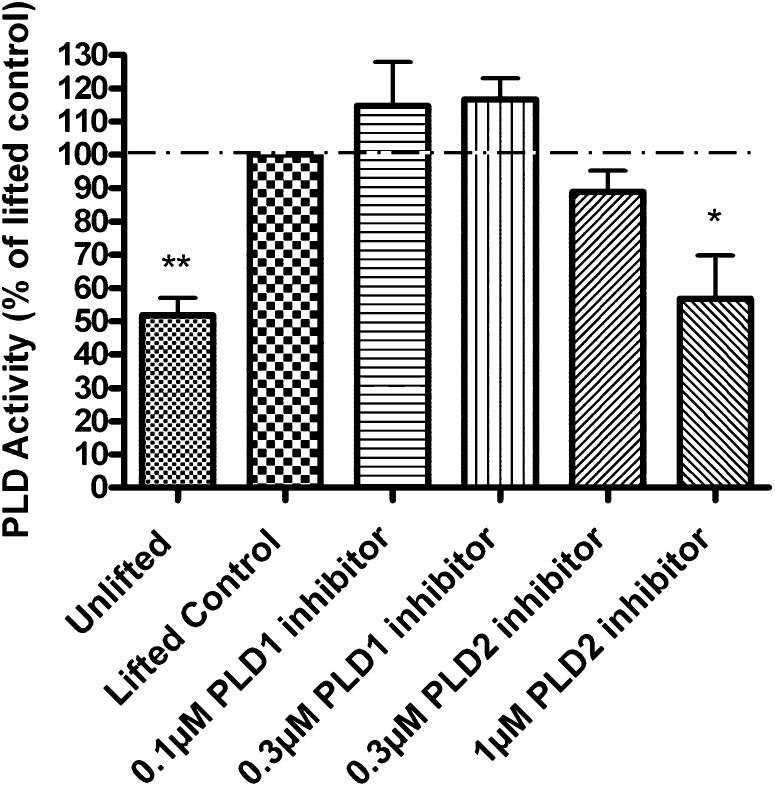
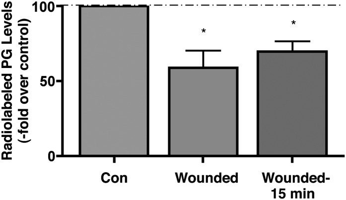
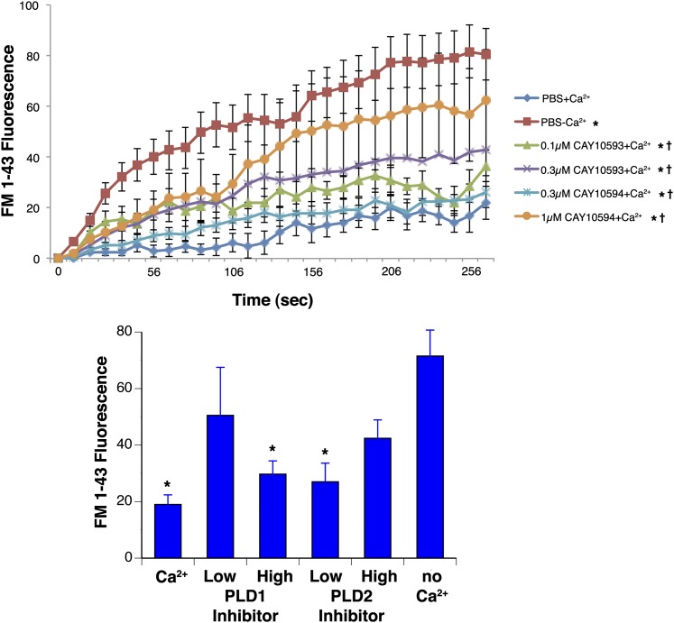
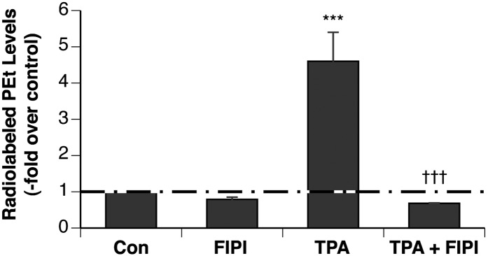
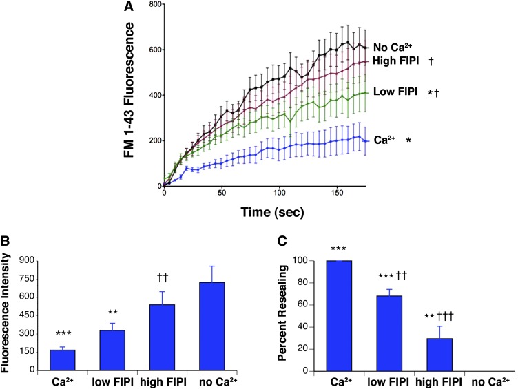
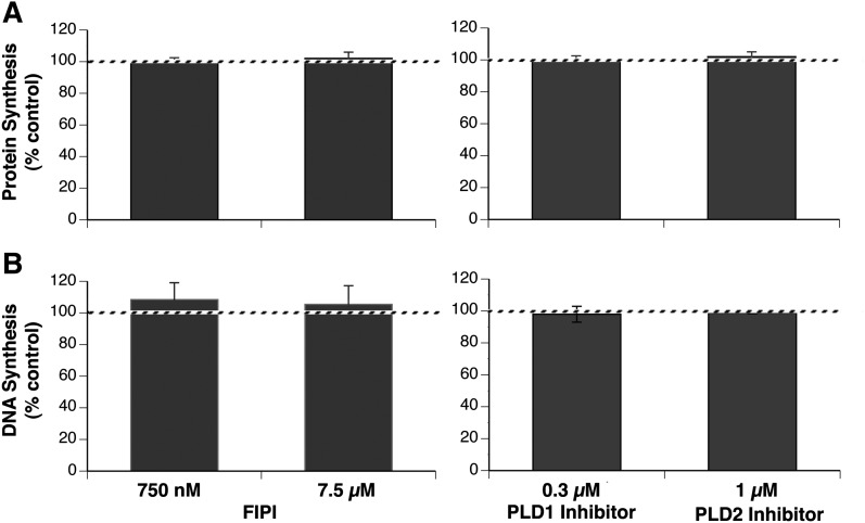
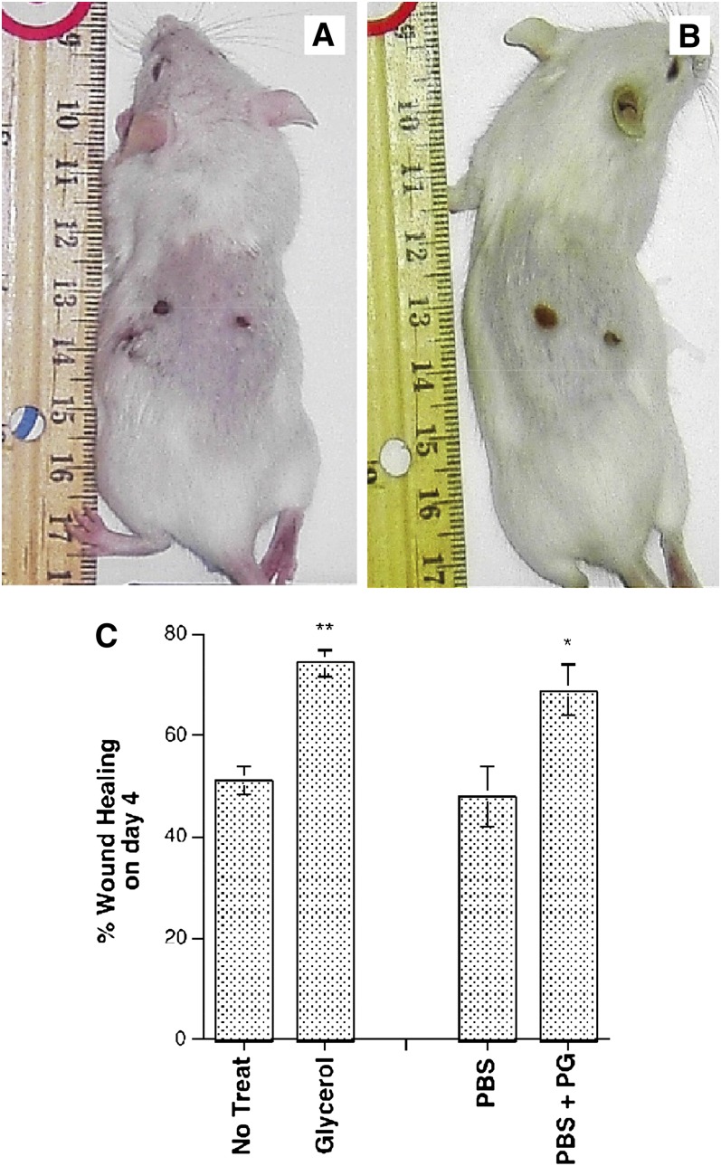
Plasma membrane disruptions occur in mechanically active tissues such as the epidermis and can lead to cell death if the damage remains unrepaired. Repair occurs through fusion of vesicle patches to the damaged membrane region. The enzyme phospholipase D (PLD) is involved in membrane traffickiing; therefore, the role of PLD in membrane repair was investigated. Generation of membrane disruptions by lifting epidermal keratinocytes from the substratum induced PLD activation, whereas removal of cells from the substratum via trypsinization had no effect. Pretreatment with 1,25-dihydroxyvitamin D₃, previously shown to increase PLD1 expression and activity, had no effect on, and a PLD2-selective (but not a PLD1-selective) inhibitor decreased, cell lifting-induced PLD activation, suggesting PLD2 as the isoform activated. PLD2 interacts functionally with the glycerol channel aquaporin-3 (AQP3) to produce phosphatidylglycerol (PG); however, wounding resulted in decreased PG production, suggesting a potential PG deficiency in wounded cells. Cell lifting-induced PLD activation was transient, consistent with a possible role in membrane repair, and PLD inhibitors inhibited membrane resealing upon laser injury. In an in vivo full-thickness mouse skin wound model, PG accelerated wound healing. These results suggest that PLD and the PLD2/AQP3 signaling module may be involved in membrane repair and wound healing.


Figures

Fig. 1.
Fig. 1.
Cell wounding, but not trypsinization, activated PLD. A: [3H]oleate-prelabeled keratinocytes in SFKM were treated with 1% ethanol immediately prior to gentle removal of the cells from the substratum with a rubber policeman or trypsinization (with 0.25% trypsin) and incubation for 15 min. Reactions were terminated by the addition of 0.2% SDS containing 5 mM EDTA, and [3H]PEt was extracted, separated by TLC, and quantified. B: [3H]oleate-prelabeled keratinocytes in HBSS were treated, and radiolabeled PEt levels were quantified as in A. Values represent the means ± SEM of three to four experiments performed in duplicate and expressed as the -fold over the control level; *P < 0.05 versus the control.
Fig. 2.
Fig. 2.
Cell wounding, but not trypsinization, activated PLD in a transient manner. [3H]oleate-prelabeled keratinocytes in SFKM were treated with 1% ethanol immediately prior to gentle removal of the cells from the substratum with a rubber policeman (wounding) or 15 min after lifting and incubation for 15 min (wounding − 15 min). Note that all conditions were incubated with 1% ethanol for 15 min. Reactions were terminated by the addition of 0.2% SDS containing 5 mM EDTA, and [3H]PEt was extracted, separated by TLC, and quantified. Values are expressed as -fold over the control and represent the means ± SEM from four separate experiments performed in duplicate; *P < 0.01 versus the control value.
Fig. 3.
Fig. 3.
Pretreatment with 1,25(OH)2D3 had no enhancing effect on PLD activation induced by cell wounding. Cells were pretreated with or without 250 nM 1,25(OH)2D3 and prelabeled with [3H]oleate for 24 h in SFKM prior to assay of PLD activity upon cell lifting as in Fig. 1. Values are expressed as -fold over the control (with or without 1,25(OH)2D3 pretreatment) and represent the means ±SEM from four separate experiments performed in duplicate; *P < 0.01 versus the control value.
Fig. 4.
Fig. 4.
A PLD2-selective but not a PLD1-selective inhibitor inhibited PLD activation induced by cell wounding. [3H]oleate-prelabeled cells were pretreated with or without the indicated concentrations of each inhibitor for 30 min in K-SFM prior to assay of PLD activity upon cell lifting as in Fig. 1. Values are expressed as -fold over the lifted control and represent the means ± SEM of three separate experiments performed in duplicate; *P < 0.05, **P < 0.01 versus the lifted control value.
Fig. 5.
Fig. 5.
Cell wounding inhibited PG production. Cells were treated with [14C]glycerol immediately prior to, or 15 minutes after, cell lifting and incubation for 15 min. Reactions were terminated by the addition of 0.2% SDS containing 5 mM EDTA, and [14H]PG was extracted, separated by TLC, and quantified. Values are expressed as -fold over the control and represent the means ± SEM of three separate experiments performed in duplicate; *P < 0.05, versus the control value. Similar results were obtained when cells were more forcefully lifted from the dish using a plastic cell lifter.
Fig. 6.
Fig. 6.
The isoform-selective PLD inhibitors tended to delay membrane repair following laser-induced membrane disruption. Keratinocytes were treated with the PLD1-selective inhibitor CAY10593 (0.1 or 0.3 µM), the PLD2-selective inhibitor CAY10594 (0.3 or 1 µM), or 0.1% DMSO (vehicle control) for approximately 30 min and then wounded using a sapphire laser at a wavelength of 835 nm. The control cells were wounded in PBS in the presence (Ca2+) and absence (no Ca2+) of calcium as positive and negative controls, respectively. The inhibitor-treated cells were wounded in PBS containing calcium. A: Shown are the means ± SEM of the fluorescence intensity of a minimum of eight cells from a representative experiment; *P < 0.05 versus the values in PBS in the absence of calcium; †P < 0.05 versus the values of PBS in the presence of calcium determined using a Tukey's multiple comparison test. B: Shown are the cumulative means ± SEM from three experiments in which the fluorescence intensity at 165 s after wounding was monitored under each condition in a minimum of eight cells; *P < 0.05 versus the values obtained in PBS lacking calcium.
Fig. 7.
Fig. 7.
The PLD inhibitor FIPI decreased PLD activity stimulated by TPA. [3H]oleate-prelabeled keratinocytes in SFKM were pretreated for 20 min with 750 nM FIPI prior to stimulation with or without 100 nM TPA in the presence of 0.5% ethanol for 30 min. Reactions were terminated by the addition of 0.2% SDS containing 5 mM EDTA, and [3H]PEt was extracted, separated by TLC, and quantified. Values represent the means ± SEM of three separate experiments performed in triplicate; ***P < 0.001 versus the control; †††P < 0.01 versus TPA alone.
Fig. 8.
Fig. 8.
The PLD inhibitor FIPI delayed membrane repair following laser-induced membrane disruption. Keratinocytes were treated with FIPI (750 nM or 7.5 µM), or 0.1% DMSO (vehicle control) for 30 min and then wounded using a sapphire laser at a wavelength of 835 nm. The control cells were wounded in PBS in the presence (Ca2+) or absence (no Ca2+) of calcium as positive and negative controls, respectively. The FIPI-treated cells were wounded in PBS containing calcium. A: Shown are the means ± SEM of the fluorescence intensity of a minimum of six cells from a representative experiment; *P < 0.05 versus the values in PBS in the absence of calcium; †P < 0.05 versus the values in PBS in the presence of calcium determined using a Tukey's multiple comparison test. B: Shown are the cumulative means ± SEM from six experiments in which the fluorescence intensity at 164 s after wounding was monitored under each condition in a minimum of six cells; *P < 0.05, **P < 0.01, ***P < 0.001 versus the values obtained in PBS lacking calcium; †P < 0.05, ††P < 0.01 versus in PBS without calcium. In panel C the data are replotted as the percentage resealing, with the recovery in the presence of calcium set to 100% and that in the absence of calcium to 0%; *P < 0.05, **P < 0.01, ***P < 0.001 versus the values obtained in PBS lacking calcium; †P < 0.05, ††P < 0.01, †††P < 0.001 versus in PBS without calcium.
Fig. 9.
Fig. 9.
The PLD inhibitors had no effect on protein or DNA synthesis, suggesting a lack of cytotoxicity. Keratinocytes were pretreated for 2 h with K-SFM containing vehicle (0.1% DMSO), 0.3 µM PLD1-selective inhibitor CAY10593, 1 µM PLD2-selective inhibitor CAY10594, or 750 nM or 7.5 µM FIPI. A: [3H]leucine or B: [3H]thymidine, at a final concentration of 1 µCi/ml, was then added for an additional 1 h. Macromolecules were precipitated by the addition of cold 5% trichloroacetic acid and, following washing, were solubilized with NaOH and counted by liquid scintillation spectroscopy. Results represent the means ± SEM of five separate experiments performed in duplicate and expressed relative to the vehicle control. The value for treatment with any of the PLD inhibitors at the indicated concentrations was not significantly different from the control.
Fig. 10.
Fig. 10.
PG liposomes accelerated wound healing of full-thickness punch biopsies of mouse skin. Two full-thickness skin punch biopsies of ∼4 mm were made on the backs of ICR CD-1 mice. For each mouse, one wound was either (A) untreated (left) or treated with 2 M glycerol (as a positive control) in water (right) or (B) treated with PBS lacking divalent cations (PBS, left) or PBS containing 100 µg/ml PG, prepared as liposomes via bath sonication (right). The rate of wound healing was then monitored. Shown is the extent of wound healing on day 4 for two representative mice. Both male and female mice were used and the results pooled. C: Two full-thickness skin punch biopsies were made as described. The rate of wound healing was then followed over 4 days by digital photography and computer image analysis, and percentage of wound healing on day 4 relative to day 1 for each of the four groups is shown. The experiment was repeated on a second group of mice, with the opposite side exposed to glycerol or PG liposomes. No difference was observed between male and female mice, so the results were pooled. Results represent the means ± SEM of eight mice for each condition; *P < 0.02 versus treatment with PBS; **P < 0.001 versus no treatment.

Similar articles

Cited by

References

    1. Bollag W. B., Dodd M. E., Shapiro B. A. 2004. Protein kinase D and keratinocyte proliferation. Drug News Perspect. 17: 117–126 - PubMed
    1. Chamcheu J. C., Siddiqui I. A., Syed D. N., Adhami V. M., Liovic M., Mukhtar H. 2011. Keratin gene mutations in disorders of human skin and its appendages. Arch. Biochem. Biophys. 508: 123–137 - PMC - PubMed
    1. McNeil P. L., Kirchhausen T. 2005. An emergency response team for membrane repair. Nat. Rev. Mol. Cell Biol. 6: 499–505 - PubMed
    1. Miyake K., Tanaka T., McNeil P. L. 2006. Disruption-induced mucus secretion: repair and protection. PLoS Biol. 4: e276. - PMC - PubMed
    1. McNeil P. L., Ito S. 1989. Gastrointestinal cell plasma membrane wounding and resealing in vivo. Gastroenterology. 96: 1238–1248 - PubMed

Publication types

LinkOut - more resources