. 2013 Jan 1;36(1):31-40.
doi: 10.5665/sleep.2296.

Effects of orexin gene transfer in the dorsolateral pons in orexin knockout mice

Affiliations

Effects of orexin gene transfer in the dorsolateral pons in orexin knockout mice

Carlos Blanco-Centurion et al. Sleep. .

Abstract

Study objectives: Narcolepsy is a sleep disorder characterized by loss of orexin neurons. Previously, our group demonstrated that transfer of the orexin gene into surrogate neurons in the lateral hypothalamus and the zona incerta significantly reduced cataplexy bouts in the orexin-ataxin-3 mice model of narcolepsy. The current study determined the effects of orexin gene transfer into the dorsolateral pontine neurons in the orexin knockout (KO) mice model of narcolepsy. The dorsolateral pons was chosen because it plays a critical role in regulating muscle tone and thus it is conceivable to be involved in cataplexy as well. Cataplexy is the pathognomonic symptom in narcolepsy.

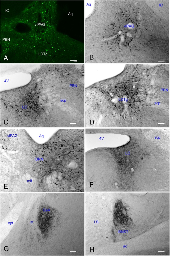
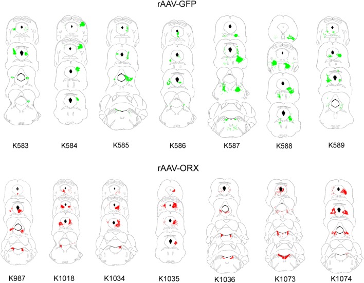
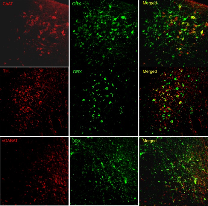
Design: Independent groups of orexin KO mice were given bilateral microinjections (0.75 μL each side) of either recombinant adenoassociated virus-orexin (rAAV-orexin; n = 7), or rAAV-green fluorescent protein (rAAV-GFP; n = 7) into the dorsolateral pons. A group of orexin KO mice that did not receive rAAV (n = 7) and a group of wild-type mice (C57BL/J6; n = 5) were used as controls. Three weeks after rAAV-mediated gene transfer narcolepsy symptoms were examined using sleep and behavioral recordings. Number, location of the orexin-immunoreactive neurons, and relative density of orexin immunoreactive fibers were determined.

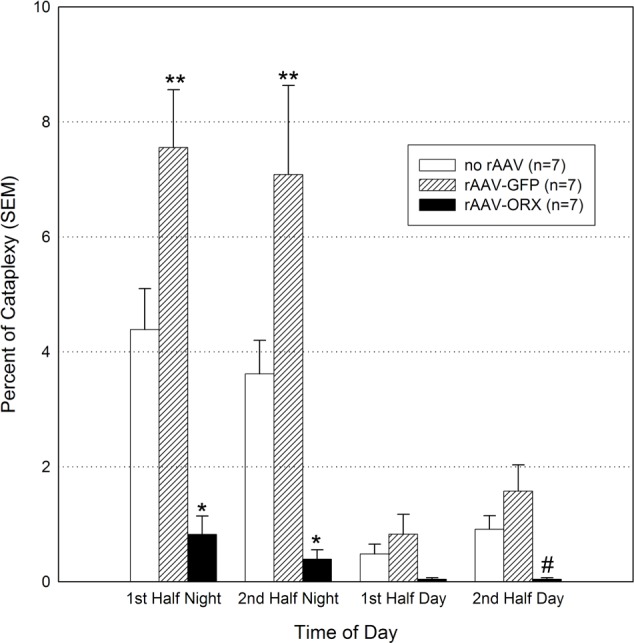
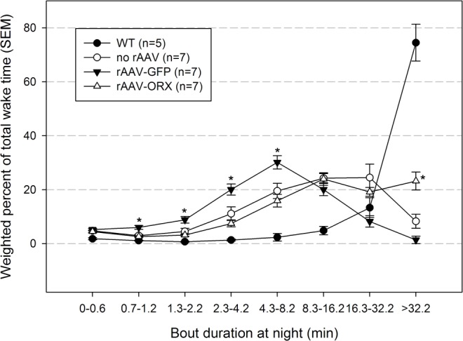
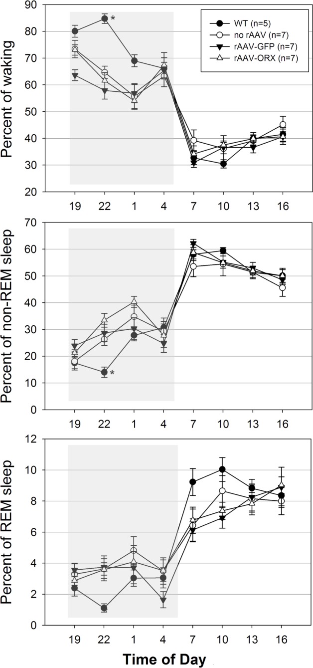
Measurements and results: Orexin gene transfer into the dorsolateral pons significantly decreased cataplexy and modestly improved wake maintenance compared to the orexin KO mice that did not receive rAAV. In contrast, GFP gene transfer worsened narcoleptic symptoms compared to the no-rAAV orexin KO group.

Conclusion: Orexin gene transfer into the dorsolateral pontine neurons can control cataplexy attacks and modestly improve wake maintenance.


Figures

Figure 1
Figure 1
Orexin gene transfer into the dorsolateral pontine neurons significantly decreases cataplexy in orexin (ORX) knockout mice. Twenty-one days after recombinant adenoassociated virus (rAAV)-mediated gene transfer sleep and behavior were recorded for 48 h. The data summarize the percent of time spent in cataplexy in 6-h bins. *P < 0.01 versus no rAAV and rAAV-green fluorescent protein (GFP); #P < 0.001 vs. rAAV-GFP; **P < 0.01 versus no rAAV. SEM, standard error of the mean.
Figure 2
Figure 2
Effects of recombinant adenoassociated virus (rAAV)-mediated gene transfer on wake maintenance during the night. Individual wake bouts were grouped into bins of different durations. Data represent group means ± standard error of the mean (SEM). Orexin (ORX) knockout mice given rAAV-orexin into the dorsolateral pons had a significant increase in the percent of time spent in long bouts. *P < 0.05 versus no rAAV. GFP, green fluorescent protein; WT, wild-type.
Figure 3
Figure 3
Effects of gene transfer on percent of sleep-wake states across 24 h in orexin (ORX) knockout and wild-type (WT) mice. Percent of time spent in vigilance states was determined 21 days after microinjection of rAAV-GFP, rAAV-ORX or no injection into the dorsolateral pons. WT (C57BL/6J) mice were also recorded and represent additional controls. Shaded boxes represent the lights-off period (19:00-07:00). *P < 0.05 versus all experimental groups. GFP, green fluorescent protein; rAAV, recombinant adenoassociated virus.
Figure 4
Figure 4
Recombinant adenoassociated viral vector (rAAV) gene transfer produced robust gene product in brains of orexin knockout mice 3 wk after its administration. Reporter gene (GFP) expression was clearly seen in somata and proximal dendrites in the dorsolateral pontine region (panel A). Robust expression of the orexin-A-ir was observed in this area including the ventrolateral periaqueductal gray (vlPAG; panel B), locus coeruleus (LC; panel C), laterodorsal tegmental nucleus (LDTg; panel D), and dorsal raphe nucleus (DRN; panel E). Orexin-A-ir was also observed in distal terminals within the LC area (Panel F), the central nucleus of the amygdala (CeA; panel G) and the bed nucleus of the stria terminalis (BNST; panel H) among others (see Table 3). White scale bars represent 100 μm. 4V, 4th ventricle; ac, anterior commissure; Aq, aquedectus of silvyus; IC, inferior colliculus, mlf, medial longitudinal fasciculus; LS, lateral septal nucleus; opt, optic tract; PBN, parabrachial nucleus; scp, superior cerebellar peduncle; st, stria terminalis.
Figure 5
Figure 5
Schematic distribution of infected neurons. All coronal sections containing cells expressing either GFP (green, top panel) or orexin-A (red, bottom panel) were plotted onto plates from Franklin and Paxinos mouse brain atlas. The numbers below each series represent individual mice. GFP, green fluorescent protein; rAAV, recombinant adenoassociated virus.
Figure 6
Figure 6
rAAV-orexin gene transfer into the dorsolateral pons produced orexin A-ir in cholinergic (top row), noradrenergic (middle row) and gamma aminobutyric acid (GABA)ergic neurons (bottom row). GABAergic neurons were labeled using a vesicular GABA transporter (vGABAT) antibody. Images were acquired using a Fluoview 1000 Olympus confocal laser scanner microscope and two excitation lines (488 and 543 nm) tuned to detect Alexa Fluor 488 (520 nm; ORX, orexin) and Alexa Fluor 568 (603 nm; ChAT, choline acetyltransferase; TH, tyrosine hydroxylase; and vGABAT, vesicular GABA transporter) dyes, respectively.

References

    1. Aldrich MS. The neurobiology of narcolepsy-cataplexy. Prog Neurobiol. 1993;41:533–41. - PubMed
    1. Overeem S, Mignot E, van Dijk JG, Lammers GJ. Narcolepsy: clinical features, new pathophysiologic insights, and future perspectives. J Clin Neurophysiol. 2001;18:78–105. - PubMed
    1. Anic-Labat S, Guilleminault C, Kraemer HC, Meehan J, Arrigoni J, Mignot E. Validation of a cataplexy questionnaire in 983 sleep-disorders patients. Sleep. 1999;22:77–87. - PubMed
    1. Krahn LE, Lymp JF, Moore WR, Slocumb N, Silber MH. Characterizing the emotions that trigger cataplexy. J Neuropsychiatry Clin Neurosci. 2005;17:45–50. - PubMed
    1. Longstreth WT, Jr, Koepsell TD, Ton TG, Hendrickson AF, van Belle G. The epidemiology of narcolepsy. Sleep. 2007;30:13–26. - PubMed

Publication types

MeSH terms