

Four novel RS1 gene mutations in Polish patients with X-linked juvenile retinoschisis
- PMID: 23288992
- PMCID: PMC3534142
Four novel RS1 gene mutations in Polish patients with X-linked juvenile retinoschisis
Abstract
Purpose: To determine the clinical features and to identify mutations in the retinoschisis gene (RS1) in ten patients with X-linked retinoschisis (XLRS).
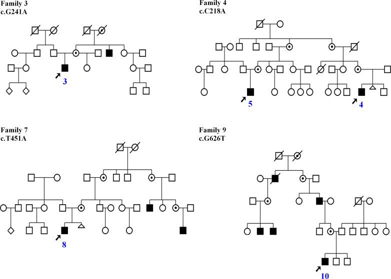
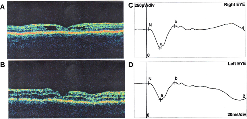
Methods: Ten male patients from nine Polish families were included in this study. Ophthalmologic examinations, including optical coherence tomography (OCT) and full-field electroretinography (ERG), were performed in all affected boys. The entire coding region encompassing six exons of the RS1 gene was amplified with PCR and directly sequenced in all the patients.
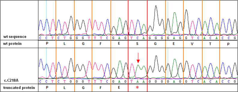
Results: All affected individuals showed typical retinoschisis signs and symptoms, and all appeared to have a mutation in the RS1 gene. Seven different mutations were identified, including two novel missense substitutions: c.176G>C (p.Cys59Ser), c.451T>A (p.Tyr151Asp); one novel nonsense substitution: c.218C>A (p.Ser73*); and one novel frameshift mutation: c.354_355delCA (p.Asp118Glufs*2). We also found two missense substitutions that had been previously described: c.214G>A (p.Glu72Lys) and c.626G>T (p.Arg209Leu) and one known splice site mutation in intron 5: c.522+1G>T (IVS5+1G>T).
Conclusions: This study provides the first molecular genetic characteristics of patients with juvenile retinoschisis from the previously unexplored Polish population. We investigated the molecular background of XLRS in ten boys. The present study reports for the first time four novel mutations, including two missense substitutions, one nonsense substitution, and one frameshift deletion. One of these substitutions and 2-bp deletion created stop codons. Moreover, we described three substitutions that had been previously reported (one is a splicing mutation). Further genetic characterization of Polish patients with XLRS will be helpful in understanding the worldwide spectrum of RS1 mutations. Despite the mutation heterogeneity found in a small group of our patients, they presented a relatively uniform clinical picture. Identifying the causative mutation is helpful in confirming diagnosis and counseling, but cannot provide prognostic data.
Figures
References
-
- Functional implications of the spectrum of mutations found in 234 cases with X-linked juvenile retinoschisis. The Retinoschisis Consortium. Hum Mol Genet. 1998;7:1185–92. - PubMed
-
- Sauer CG, Gehrig A, Warneke-Wittstock R, Marquardt A, Ewing CC, Gibson A, Lorenz B, Jurklies B, Weber BH. Positional cloning of the gene associated with X-linked juvenile retinoschisis. Nat Genet. 1997;17:164–70. - PubMed
-
- Grayson C, Reid SN, Ellis JA, Rutherford A, Sowden JC, Yates JR, Farber DB, Trump D. Retinoschisin, the X-linked retinoschisis protein, is a secreted photoreceptor protein, and is expressed and released by Weri-Rb1 cells. Hum Mol Genet. 2000;9:1873–9. - PubMed
-
- Molday LL, Hicks D, Sauer CG, Weber BH, Molday RS. Expression of X-linked retinoschisis protein RS1 in photoreceptor and bipolar cells. Invest Ophthalmol Vis Sci. 2001;42:816–25. - PubMed
Publication types
MeSH terms
Substances
LinkOut - more resources
Full Text Sources