

Identifying targets of the Sox domain protein Dichaete in the Drosophila CNS via targeted expression of dominant negative proteins
- PMID: 23289785
- PMCID: PMC3541953
- DOI: 10.1186/1471-213X-13-1
Identifying targets of the Sox domain protein Dichaete in the Drosophila CNS via targeted expression of dominant negative proteins
Abstract
Background: Group B Sox domain transcription factors play important roles in metazoan central nervous system development. They are, however, difficult to study as mutations often have pleiotropic effects and other Sox family members can mask phenotypes due to functional compensation. In Drosophila melanogaster, the Sox gene Dichaete is dynamically expressed in the embryonic CNS, where it is known to have functional roles in neuroblasts and the ventral midline. In this study, we use inducible dominant negative proteins in combination with ChIP, immunohistochemistry and genome-wide expression profiling to further dissect the role of Dichaete in these two tissues.
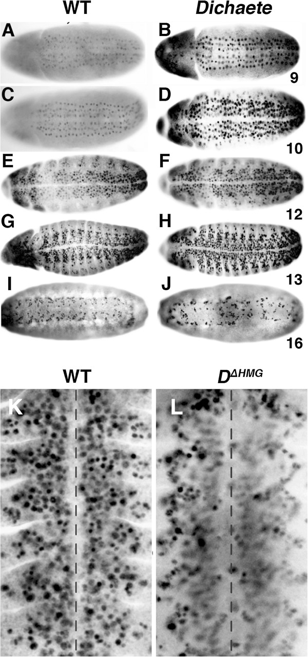
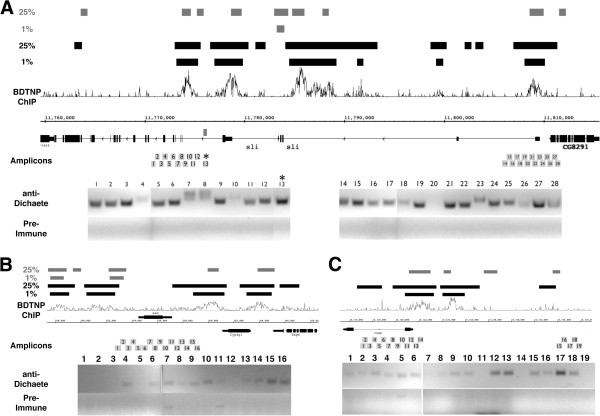
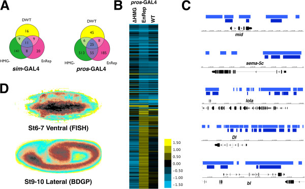
Results: We generated two dominant negative Dichaete constructs, one lacking a DNA binding domain and the other fused to the Engrailed transcriptional repressor domain. We expressed these tissue-specifically in the midline and in neuroblasts using the UAS/GAL4 system, validating their use at the phenotypic level and with known target genes. Using ChIP and immunohistochemistry, we identified two new likely direct Dichaete target genes, commisureless in the midline and asense in the neuroectoderm. We performed genome-wide expression profiling in stage 8-9 embryos, identifying almost a thousand potential tissue-specific Dichaete targets, with half of these genes showing evidence of Dichaete binding in vivo. These include a number of genes with known roles in CNS development, including several components of the Notch, Wnt and EGFR signalling pathways.
Conclusions: As well as identifying commisureless as a target, our data indicate that Dichaete helps establish its expression during early midline development but has less effect on its established later expression, highlighting Dichaete action on tissue specific enhancers. An analysis of the broader range of candidate Dichaete targets indicates that Dichaete plays diverse roles in CNS development, with the 500 or so Dichaete-bound putative targets including a number of transcription factors, signalling pathway components and terminal differentiation genes. In the early neurectoderm we implicate Dichaete in the lateral inhibition pathway and show that Dichaete acts to repress the proneural gene asense. Our analysis also reveals that dominant negatives cause off-target effects, highlighting the need to use other experimental data for validating findings from dominant negative studies.
Figures
References
-
- Kalinka AT, Varga KM, Gerrard DT, Preibisch S, Corcoran DL, Jarrells J, Ohler U, Bergman CM, Tomancak P. Gene expression divergence recapitulates the developmental hourglass model. Nature. 2010;468:811–814. - PubMed
-
- Masui S, Nakatake Y, Toyooka Y, Shimosato D, Yagi R, Takahashi K, Okochi H, Okuda A, Matoba R, Sharov AA. et al.Pluripotency governed by Sox2 via regulation of Oct3/4 expression in mouse embryonic stem cells. Nat Cell Biol. 2007;9(6):625–635. - PubMed
-
- Blanco J, Girard F, Kamachi Y, Kondoh H, Gehring WJ. Functional analysis of the chicken delta1-crystallin enhancer activity in Drosophila reveals remarkable evolutionary conservation between chicken and fly. Development. 2005;132:1895–1905. - PubMed
-
- Nambu P, Nambu J. The Drosophila fishhook gene encodes a HMG domain protein essential for segmentation and CNS development. Development. 1996;122:3467–3475. - PubMed
-
- Russell SRH, Sanchez-Soriano N, Wright CR, Ashburner M. The Dichaete gene of Drosophila melanogaster encodes a SOX-domain protein required for embryonic segmentation. Development. 1996;122:3669–3676. - PubMed
Publication types
MeSH terms
Substances
Grants and funding
LinkOut - more resources
Full Text Sources
Other Literature Sources
Molecular Biology Databases
Research Materials
Miscellaneous
