. 2013 Mar 1;437(1):12-9.
doi: 10.1016/j.virol.2012.12.004. Epub 2013 Jan 4.

Human papillomavirus infection is inhibited by host autophagy in primary human keratinocytes

Affiliations

Human papillomavirus infection is inhibited by host autophagy in primary human keratinocytes

Laura M Griffin et al. Virology. .

Abstract

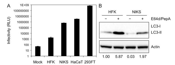
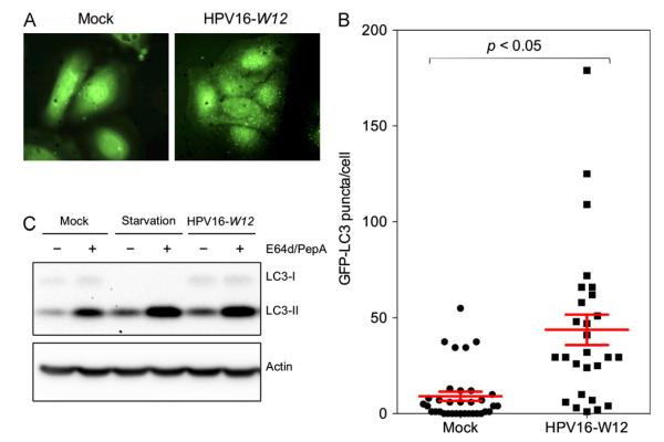
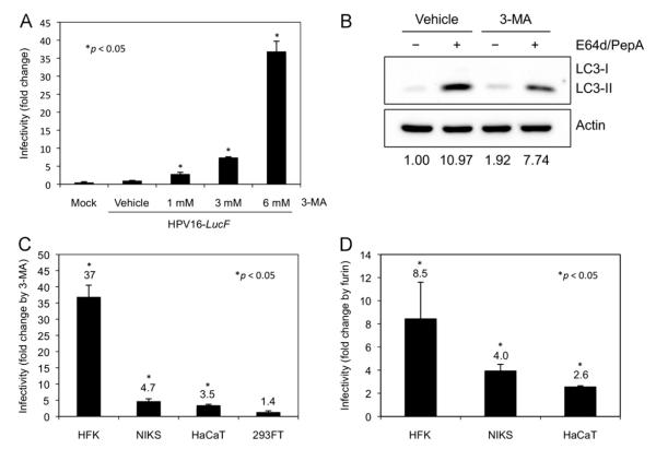
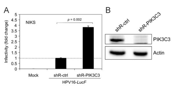
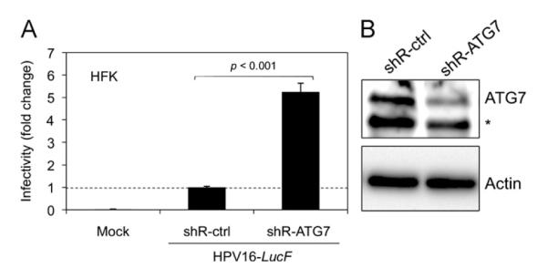
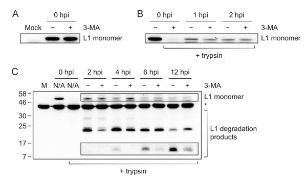
Human papillomavirus (HPV) infection is severely limited in its natural host, primary human keratinocytes. Our data show HPV infectivity in primary keratinocytes is over 100- and 1,000-fold lower than in established keratinocyte cell lines NIKS and HaCaT, respectively. Here, we show that the basal level of autophagy in primary human foreskin keratinocytes (HFKs) is higher than in immortalized keratinocytes, and that HPV16 virions significantly induce autophagy in HFKs. Interestingly, HPV16 infectivity is dramatically enhanced by knockdown of essential autophagy genes as well as biochemical inhibition of autophagy. The increase in HPV16 infectivity by autophagy inhibition is most significant in HFKs, showing an inverse correlation with basal HPV16 infectivity in HFK, NIKS, HaCaT, and 293FT cells. Further, inhibition of autophagy delays degradation of HPV16 capsid proteins during virus trafficking, indicating that host autophagy induced by HPV16 virions inhibits infection of primary keratinocytes through rapid degradation of viral capsid proteins.


Figures

Fig. 1
Fig. 1. Primary keratinocytes show limited HPV16 infectivity and high levels of basal autophagy
(A) The indicated cell lines were inoculated with 10,000 vge/cell of HPV16-LucF and incubated for 48 h. HPV16 infectivity and cell viability were measured by Bright-GloTM Luciferase Assay System (Promega)and CellTiter-Glo Luminescent Cell Vi ability Assay (Promega), respectively. Infectivity data normalized to cell viability are shown as average relative luminescence units (RLU) from quadruplicate samples. The data shown here are from one representative of three independent experiments. (B) HFK and NIKS cells were treated with the protease inhibitors E64d and Pepstatin A (10 μg/mL each) or DMSO for 6 h, and harvested for immunoblotting of LC3, using Actin as an internal control.The data shown here are from one representative of three independent experiments.
Fig. 2
Fig. 2. HPV16 virions induce host autophagy in primary keratinocytes
(A) HFKs were transduced with GFP-LC3-containing lentiviruses for 48 h, inoculated with 1,000 vge/cell of HPV16-W12, and incubated for 6 h. Cells were fixed with 4% paraformaldehyde and mounted with ProLong® Gold Antifade Reagent with DAPI (Life Technologies). At least 25 cells were imaged for each condition. Representative images are shown. (B) Quantification of GFP-LC3 puncta obtained in (A). GFP-LC3 puncta were counted using NIH ImageJ image analysis software and plotted to show the number of GFP-LC3 puncta per cell. One-way analysis of variance between groups (ANOVA) statistical analysis to determine the p-value was performed using GraphPad Prism software (GraphPad Software, La Jolla, CA). Data shown are from one representative of two independent experiments. (C) HFKs were incubated with Krebs–Ringer Bicarbonate buffer for starvation condition, or 1,000 vge/cell of HPV16-W12 for 6 h. LC3 and Actin were detected by immunoblotting. The data shown here are from one representative of three independent experiments.
Fig. 3
Fig. 3. Inhibition of autophagy by 3-MA significantly enhances HPV16 infectivity in primary keratinocytes
(A) Following pretreatment with the indicated concentrations of 3-MA or vehicle for 4 h, HFKs were inoculatedwith 10,000 vge/cell of HPV16-LucF and incubated for 48 h in the presence of 3-MA or vehicle. Infectivity and cell viability were measured as described above. Normalized infectivity data are presented as fold change in infectivity by 3-MA over vehicle control, and were averaged from quadruplicate samples. (B) HFKs were treated with 6 mM 3-MA or vehicle for 6 h with protease inhibitors E64d and Pepstatin A (10 μg/mL each) or DMSO, and harvested for immunoblotting of LC3 and Actin. (C) The indicated cell lines were treated with 6 mM 3-MA or vehicle, and inoculated with HPV16-LucF as in (A). Normalized infectivity data are shown as fold changein infectivity by 3-MA over vehicle control. (D) The indicated cell lines were treated with 8 U/mL furin or vehicle, and inoculated with HPV16-LucF as in (A). Normalized infectivity data are shown as fold change in infectivity by furin over vehicle control. The data shown here are from one representative of three independent experiments. Student’s t-test was performed to determine p-values between 3-MA treatments and vehicle control using StatPlus® (AnalystSoft, Inc.).
Fig. 4
Fig. 4. HPV16 infectivity is increased by knockdown of PIK3C3
(A) NIKS cells were transduced with lentiviruses containing shRNAs against PIK3C3 (shR-PIK3C3) or a non-target control (shR-ctrl). At 48 h post transduction, the transduced cells were selected by culture in medium containing 1.5 μg/mL of puromycin for 2 weeks to establish stable semi-clonal cell lines. Puromycin-selected cells were inoculated with 10,000 vge/cell of HPV16-LucF and incubated for 48 h. Infectivity was measured and normalized to cell viability as described in Fig. 1. Normalized infectivity data are shown as fold change in infectivity by shR-PIK3C3 over shR-ctrl, and were averaged from quadruplicate samples. Statistical analysis was performed as described in Fig. 3A. (B) In parallel to the infection assay in (A), immunoblotting was performed with the puromycin-selected cells using anti-PIK3C3 (Life Technologies) and anti-Actin antibodies. The data shown here are from one representative of three independent experiments.
Fig. 5
Fig. 5. HPV16 infectivity is increased by knockdown of the essential autophagy gene ATG7
(A) HFKs were transduced with lentiviruses containing shRNAs against ATG7 (shR-ATG7) or shR-ctrl and puromycin-selected as in Fig. 3A. Infection assay and analysis of puromycin-selected cells was conducted as in Fig. 3A, and data are shown as fold change in infectivity by shR-ATG7 over shR-ctrl. Statistical analysis was performed as described in Fig. 3A. (B) In parallel to the infection assay in (A), immunoblotting was performed using anti-ATG7 antibody (ProSci) and Actin. The data shown here are from one representative of three independent experiments. *Non-specific band.
Fig. 6
Fig. 6. The autophagy inhibitor, 3-MA, delays degradation of HPV16 L1 capsid proteins during entry
(A) Effect of 3-MA on virus binding. HFKs were pretreated with 6 mM 3-MA for 4 h. Cells were chilled to 4 °C to inhibit endocytosis, inoculated with 10,000 vge/cell of HPV16-W12 and incubated with or without 3-MA for 1 h at 4 °C. Unbound virions were washed out and cells were lysed directly to observe total bound virions, followed by immunoblotting of L1 capsid protein (CAMVIR-1, Abcam). (B) Effect of 3-MA on virus internalization. Virus internalization was initiated by shifting cells treated as in (A) to 37 °C. At the indicated hours post infection (hpi), cells were trypsinized prior to lysis to observe only internalized virions, followed by immunoblotting of L1 capsid protein. (C) Effect of 3-MA on capsid protein degradation during entry. Experiment was conducted as in (A) and (B) except 3-MA was added only when cells were shifted to 37 °C to initiate internalization (no pretreatment). The data shown here are from one representative of three independent experiments. Boxed bands are from a longer exposure. M, mock; N/A, not applicable. *Non-specific band.

References

    1. Abban CY, Bradbury NA, Meneses PI. HPV16 and BPV1 infection can be blocked by the dynamin inhibitor dynasore. Am. J. Ther. 2008;15:304–311. - PMC - PubMed
    1. Allen-Hoffmann BL, Schlosser SJ, Ivarie CA, Sattler CA, Meisner LF, O’Connor SL. Normal growth and differentiation in a spontaneously immortalized near-diploid human keratinocyte cell line, NIKS. J. Invest. Dermatol. 2000;114:444–455. - PubMed
    1. Blommaart EF, Krause U, Schellens JP, Vreeling-Sindelárová H, Meijer AJ. The phosphatidylinositol 3-kinase inhibitors wortmannin and LY294002 inhibit autophagy in isolated rat hepatocytes. Eur. J. Biochem. 1997;243:240–246. - PubMed
    1. Bousarghin L, Touzé A, Sizaret P-Y, Coursaget P. Human papillomavirus types 16, 31, and 58 use different endocytosis pathways to enter cells. J. Virol. 2003;77:3846–3850. - PMC - PubMed
    1. Buck CB, Pastrana DV, Lowy DR, Schiller JT. Efficient intracellular assembly of papillomaviral vectors. J. Virol. 2004;78:751–757. - PMC - PubMed

MeSH terms