. 2013 Jan 4;4(1):1.
doi: 10.1186/scrt149.

A new platelet cryoprecipitate glue promoting bone formation after ectopic mesenchymal stromal cell-loaded biomaterial implantation in nude mice

A new platelet cryoprecipitate glue promoting bone formation after ectopic mesenchymal stromal cell-loaded biomaterial implantation in nude mice

Marina Trouillas et al. Stem Cell Res Ther. .

Abstract

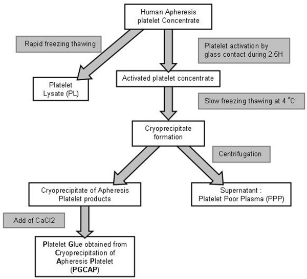
Introduction: This study investigated the promising effect of a new Platelet Glue obtained from Cryoprecipitation of Apheresis Platelet products (PGCAP) used in combination with Mesenchymal Stromal Cells (MSC) loaded on ceramic biomaterials to provide novel strategies enhancing bone repair.

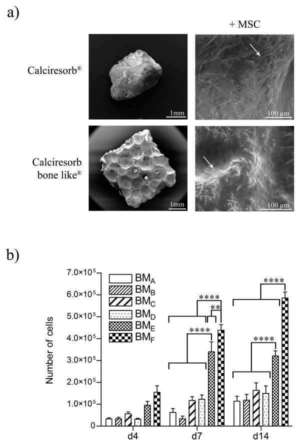
Methods: PGCAP growth factor content was analyzed by ELISA and compared to other platelet and plasma-derived products. MSC loaded on biomaterials (65% hydroxyapatite/35% beta-TCP or 100% beta-TCP) were embedded in PGCAP and grown in presence or not of osteogenic induction medium for 21 days. Biomaterials were then implanted subcutaneously in immunodeficient mice for 28 days. Effect of PGCAP on MSC was evaluated in vitro by proliferation and osteoblastic gene expression analysis and in vivo by histology and immunohistochemistry.

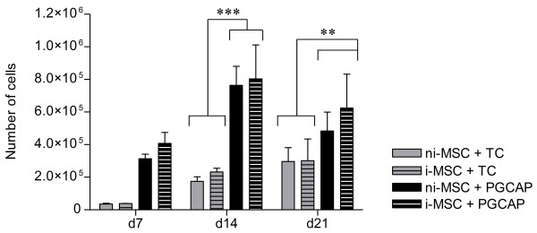
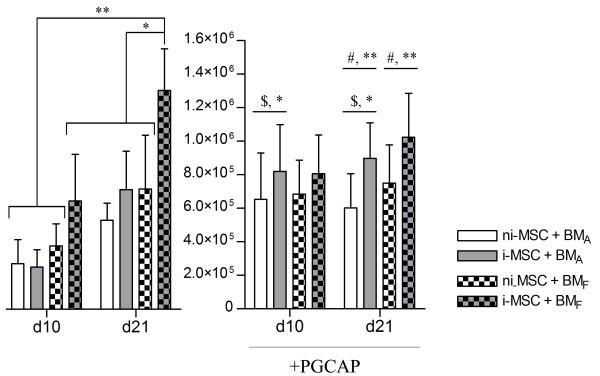
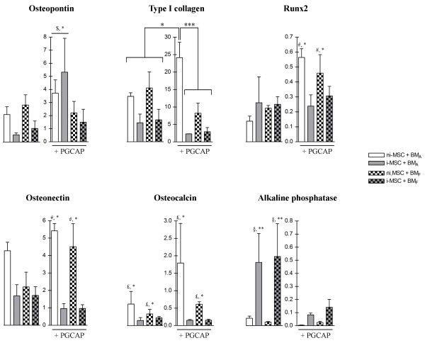
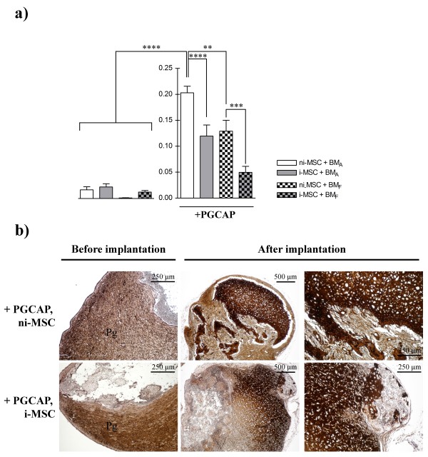
Results: We showed that PGCAP, compared to other platelet-derived products, allowed concentrating large amount of growth factors and cytokines which promoted MSC and osteoprogenitor proliferation. Next, we found that PGCAP improves the proliferation of MSC and osteogenic-induced MSC. Furthermore, we demonstrated that PGCAP up-regulates the mRNA expression of osteogenic markers (Collagen type I, Osteonectin, Osteopontin and Runx2). In vivo, type I collagen expressed in ectopic bone-like tissue was highly enhanced in biomaterials embedded in PGCAP in the absence of osteogenic pre-induction. Better results were obtained with 65% hydroxyapatite/35% beta-TCP biomaterials as compared to 100% beta-TCP.

Conclusions: We have demonstrated that PGCAP is able to enhance in vitro MSC proliferation, osteoblastic differentiation and in vivo bone formation in the absence of osteogenic pre-induction. This clinically adaptable platelet glue could be of interest for improving bone repair.


Figures

Figure 1
Figure 1
Preparation of platelet glue obtained from cryoprecipitation of apheresis platelets.
Figure 2
Figure 2
PGCAP promotes mesenchymal stromal cell (MSC) and osteoprogenitor proliferation. Not induced-MSCs (ni-MSCs) or osteogenic induced-MSCs (i-MSCs) were embedded in TC or PGCAP and grown for 21 days. The number of cells was evaluated in each condition at days 7, 10, and 21. Data are expressed as mean ± standard error of the mean. Analysis-of-variance test followed by Student Newman-Keuls test revealed statistically significant difference between groups (PGCAP and TC) at different times. **P <0.01, ***P <0.001. d, day; PGCAP, platelet glue obtained from cryoprecipitation of apheresis platelet products; TC, Tissucol.
Figure 3
Figure 3
Physical characteristics and biocompatibility of biomaterials and mesenchymal stromal cell (MSC) proliferation on the different biomaterials. (a) Representative scanning electron microscopy images of 'Calciresorb' (BMA) and 'Calciresorb bone like' (BMF) biomaterial surfaces are shown. MSCs were loaded on these two biomaterials and grown in platelet lysate (PL) medium. MSC colonization on the various biomaterials was evaluated by scanning electron microscopy at 14 days of culture. Arrows indicate cells on biomaterials. (b) MSCs loaded on the various biomaterials at low cell seeding density (100 × 103 cells per biomaterial) were grown in PL medium for 14 days. The number of cells on the different biomaterials was evaluated at days 4, 7, and 14. The data are expressed as mean ± standard error of the mean. A statistically significant difference between individual conditions was revealed by analysis-of-variance test followed by Student Newman-Keuls test. **P <0.01, ****P <0.0001. d, day.
Figure 4
Figure 4
Effect of PGCAP on mesenchymal stromal cell (MSC) and osteoprogenitor proliferation loaded on BMA and BMF. Not induced-MSCs (ni-MSCs) or osteogenic induced-MSCs (i-MSCs) loaded on BMA and BMF biomaterials at high cell seeding density (20 × 103 cells per biomaterial) were embedded or not in PGCAP and grown for 21 days. The number of cells was evaluated in each condition at days 10 and 21. The data are expressed as mean ± standard error of the mean. A statistically significant difference between individual conditions (without PGCAP) was revealed by analysis-of-variance test followed by Student Newman-Keuls. *P <0.05, **P <0.01. With PGCAP, similar tests revealed statistically significant difference between group comparing ni-MSCs and i-MSCs loaded on BMA ($*P <0.05) or group comparing ni-MSCs and i-MSCs at day 21 (#**P <0.01). PGCAP, platelet glue obtained from cryoprecipitation of apheresis platelet products.
Figure 5
Figure 5
PGCAP improves osteogenic differentiation of mesenchymal stromal cells (MSCs) loaded on biomaterials. Total RNAs were extracted from ni-MSCs and i-MSCs loaded on BMA and BMF at high cell seeding density (200 × 103 cells per biomaterial) embedded or not in PGCAP and grown for 21 days. Quantifications of osteopontin, type I collagen, Runx2, osteonectin, osteocalcin, and alkaline phosphatase mRNA expression were analyzed by real-time quantitative-polymerase chain reaction. Transcript levels in arbitrary units are expressed as mean ± standard error of the mean. A statistically significant difference between individual conditions or groups of condition was revealed by analysis-of-variance test followed by Student Newman-Keuls test. Osteopontin: group of ni-MSCs and i-MSCs loaded on BMA embedded in PGCAP was compared with other conditions ($*P <0.05). Type I collagen: individual comparisons, *P <0.05, **P <0.01, ***P <0.001. Runx2, Osteonectin: group with ni-MSCs embedded in PGCAP (loaded in both biomaterials) was compared with other conditions with i-MSCs (#*P <0.05). Osteocalcin: comparison of group osteogenic-induced or not (£*P <0.05). Alkaline phosphatase: group with i-MSCs without PGCAP (loaded in both biomaterials) was compared with other conditions with ni-MSCs (§**P <0.01). i-MSC, not osteogenic induced-mesenchymal stromal cell; ni-MSC, not induced-mesenchymal stromal cell; PGCAP, platelet glue obtained from cryoprecipitation of apheresis platelet products.
Figure 6
Figure 6
Ex vivo confocal microscopy analysis of BMA and BMF biomaterials. Not induced- mesenchymal stromal cells (ni-MSCs) or osteogenic induced-MSCs (i-MSCs) GFP+ loaded on BMA and BMF at high cell seeding density (200 × 103 cells per biomaterial) were embedded or not in PGCAP and grown for 21 days. Next, biomaterials were subcutaneously grafted on nude mice. Unloaded biomaterials (no pre-loaded cells and not embedded in PGCAP) were also grafted on mice. Twenty-eight days after grafting, biomaterials were cut in four pieces, fixed, and stained with CD90 antibody. Biomaterials were analyzed by confocal microscopy (blue: biomaterial; green: MSC GFP+; red: CD90APC; and purple: DAPI). Original magnification: ×20. Each picture is representative of three independent experiments. DAPI, 4'-6-diamidino-2-phenylindole; PGCAP, platelet glue obtained from cryoprecipitation of apheresis platelet products.
Figure 7
Figure 7
Ectopic implantation of MSC-loaded BMA embedded in PGCAP promotes bone formation in nude mice. Not induced-MSCs (ni-MSCs) or osteogenic induced-MSCs (i-MSCs) GFP+ loaded on BMA at high cell seeding density (200 × 103 cells per biomaterial) were embedded or not in PGCAP and grown for 21 days. Next, biomaterials were grafted on nude mice. After 21 days of in vitro culture (before implantation) and 28 days after grafting (after implantation), biomaterials were decalcified, embedded in paraffin, and stained with hematoxylin (nuclei), phloxin (cytoplasm), and safranin (matrix). Original magnification: ×10. Boxed area is magnified and points out cells that have migrated in biomaterials or area of bone formation. Arrows represent bone-like tissue characterized by osteocyte-like cells embedded in matrix stained by safranin. Each picture is representative of three independent experiments. B, biomaterial; F, fibroid tissue; Pg, platelet glue obtained from cryoprecipitation of apheresis platelet products; PGCAP, platelet glue obtained from cryoprecipitation of apheresis platelet products; V, vessels.
Figure 8
Figure 8
Bone formation areas are increased after ectopic implantation of MSC-loaded BMA embedded in PGCAP. (a) The average ratio of bone formation area in BMA 28 days after grafting was determined in each condition. The data are expressed as mean ± standard error of the mean. A statistically significant difference between individual conditions was revealed by analysis-of-variance test followed by Student Newman-Keuls test. *P <0.05, **P <0.01, ***P <0.00, ****P <0.0001. (b) Type I collagen immuno-staining (brownish color) was performed on BMA and BMF sections before implantation and 28 days after implantation. Original magnifications: ×4 and ×10. i-MSC, not osteogenic induced-mesenchymal stromal cell; MSC, mesenchymal stromal cell; ni-MSC, not induced-mesenchymal stromal cell; PGCAP, platelet glue obtained from cryoprecipitation of apheresis platelet products.

Similar articles

Cited by

References

    1. Cancedda R, Giannoni P, Mastrogiacomo M. A tissue engineering approach to bone repair in large animal models and in clinical practice. Biomaterials. 2007;28:4240–4250. doi: 10.1016/j.biomaterials.2007.06.023. - DOI - PubMed
    1. Heiple KG, Goldberg VM, Powell AE, Bos GD, Zika JM. Biology of cancellous bone grafts. Orthop Clin North Am. 1987;18:179–185. - PubMed
    1. Drosse I, Volkmer E, Capanna R, De Biase P, Mutschler W, Schieker M. Tissue engineering for bone defect healing: an update on a multi-component approach. Injury. 2008;39(Suppl 2):S9–20. - PubMed
    1. Dawson E, Mapili G, Erickson K, Taqvi S, Roy K. Biomaterials for stem cell differentiation. Adv Drug Deliv Rev. 2008;60:215–228. doi: 10.1016/j.addr.2007.08.037. - DOI - PubMed
    1. Chai YC, Carlier A, Bolander J, Roberts SJ, Geris L, Schrooten J, Van Oosterwyck H, Luyten FP. Current views on calcium phosphate osteogenicity and the translation into effective bone regeneration strategies. Acta Biomater. 2012;8:3876–3887. doi: 10.1016/j.actbio.2012.07.002. - DOI - PubMed

MeSH terms

LinkOut - more resources