. 2013 Jan 21;23(2):118-26.
doi: 10.1016/j.cub.2012.11.036. Epub 2013 Jan 3.

Mechanism and regulation of Cdc25/Twine protein destruction in embryonic cell-cycle remodeling

Affiliations

Mechanism and regulation of Cdc25/Twine protein destruction in embryonic cell-cycle remodeling

Jeffrey A Farrell et al. Curr Biol. .

Abstract

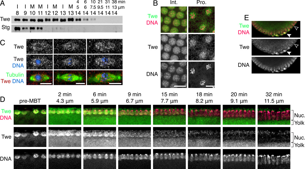
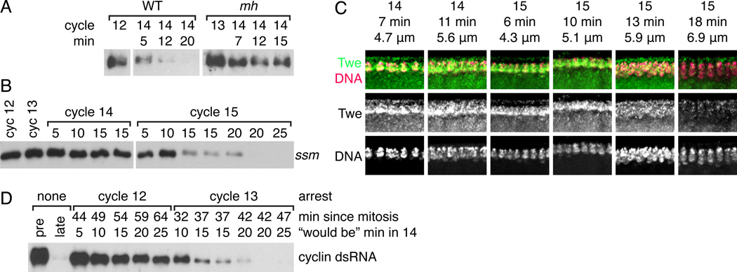
Background: In Drosophila embryos, the midblastula transition (MBT) dramatically remodels the cell cycle during the 14(th) interphase. Before the MBT, each cycle is composed of only a short S phase and mitosis. At the MBT, S phase is dramatically lengthened by the onset of late replication, and a G2 phase is introduced. Both changes set the stage for gastrulation and require downregulation of Cdc25 phosphatase, which was previously attributed to the elimination of its transcripts at the MBT.

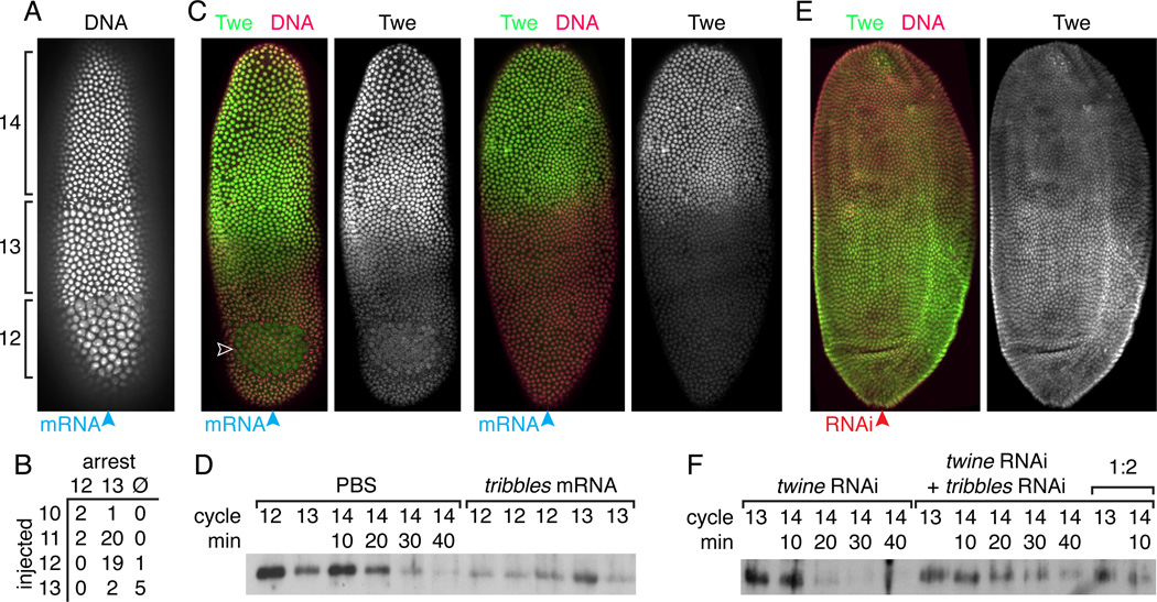
Results: Premature removal of cdc25 transcripts by RNAi did not affect progression to the MBT. Instead, an antibody against the Cdc25 isoform Twine showed that Twine protein was abundant and stable until the MBT, when it was destabilized and rapidly eliminated. Persistence of pre-MBT levels of Twine was sufficient to prevent cell-cycle slowing. Twine protein destruction was timed by the nucleocytoplasmic ratio and depended on the activation of zygotic transcription at the MBT, including expression of the gene tribbles, whose activity was sufficient to trigger Twine destruction and was required for prompt Twine disappearance.

Conclusions: We propose that the developmentally regulated destruction of Twine protein is a critical switch that contributes to the cell-cycle change at the MBT, including the addition of a G2 phase and onset of late replication. Moreover, we show that this destruction is triggered by the nucleocytoplasmic ratio-dependent onset of zygotic transcription of tribbles and other unknown genes.


Figures

Figure 1
Figure 1. RNAi against string and twine does not lengthen the cell cycle
(A) RT-PCR of string and twine in embryos injected with combined dsRNA against string and twine shows that the dsRNA triggers near complete destruction of these transcripts by 25 minutes after injection. (B) The length of interphases 12 and 13 was measured by H2AvD-GFP in uninjected embryos and embryos injected with dsRNA against string and twine 45 minutes before cycle 12.
Figure 2
Figure 2. Twine is destroyed in cycle 14, later than String
(A) Western blot of single embryos showing the destruction of String (Stg) and Twine (Twe). Embryos are labeled with their cycle number (determined by nuclear spacing) and either cell cycle stage (I = interphase, M = mitosis) or nuclear length (µm) and calculated time (min) if they are in cycle 14. (B) Twine localization during interphase and prophase. DNA (Pico Green dye, magenta) and Twine (anti-Twe, green). (C) Twine enrichment on the spindle during metaphase and anaphase. Tubulin (anti-Tubulin, green), Twine (anti-Twine, red), and DNA (Hoecsht, blue). (D) Twine destruction over the course of cycle 14. Twine (anti-Twe, green) and DNA (Pico Green dye, magenta). Micrographs are labeled with nuclear length (µm) and calculated time in cycle 14 (min). Staining of Twine is the signal that colocalizes with nuclei, whereas signal below the nuclei is fluorescence of the yolk. (E) Twine is destroyed later in the pole cells (solid arrowhead) than somatic cells (open arrowhead). Twine (anti-Twe, shown in green), DNA (Pico Green, shown in magenta).
Figure 3
Figure 3. Twine protein is relatively stable until the MBT, and pre-MBT Twine level causes a penetrant phenotype
(A–C) Western blots of embryos treated with twine RNAi, showing that Twine protein level does not depend on continued translation of its mRNA. Controls: a pre-MBT embryo (“pre”) and a 1:2 dilution (“1/2 pre”), an embryo in cycle 14 after Twine destruction (“late”). Embryos were injected at different times, aged for the time marked above each lane, and picked in cycle 11 (A), cycle 12 (B), or cycle 13 (C), as gauged by nuclear distance. (D–E) Blots showing stability of Twine protein after injection with cycloheximide (CHX), an inhibitor of translation. Dilutions of an uninjected embryo are shown (“1:1”, “1:2”, and “1:4”). Embryos were injected with CHX in cycle 12 (D) or cycle 13 (E) (and thereby arrested in that cycle) and aged for the number of minutes indicated above the lane, before being picked. (F) H2AvD-GFP embryos were injected in one end with twine mRNA at varying concentrations, and imaged to determine how many embryos underwent a shortened S phase or G2 as a result. (G) Western blot of Twine in embryos either before the MBT (“pre-MBT”) or 15 minutes into cycle 14, injected with twine mRNA at varying concentrations.
Figure 4
Figure 4. The onset of Twine destruction responds to the nucleo-cytoplasmic ratio
(A) Western blot comparing destruction of Twine in Sevelen (WT) embryos and maternal haploid embryos (mh). The cycle and time in cycle 14 (determined visually on the microscope) is marked above each lane. (B) Western blot showing destruction of Twine in haploid ssm embryos. Time in cycle 14 or 15 is marked above each lane. (C) Immunofluoresence showing Twine destruction in cycle 15 in ssm embryos. Twine (anti-Twe, green) and DNA (Pico Green dye, magenta). Micrographs are labeled with cycle, nuclear length (µm) and calculated time in cycle 14 or 15 (min) (see Fig. S2). Twine staining is signal that colocalizes with nuclei, whereas signal below the nuclei is fluorescence from the yolk. (D) Western blot showing Twine stability in embryos arrested with dsRNA against the mitotic cyclins. Lanes are labeled from top to bottom with: the cycle in which the embryo arrested, the number of minutes since it entered that cycle, and finally a calculated time, describing how far into the cycle 14 the embryo would be if it had not been arrested. Controls: a pre-MBT embryo (“pre”) and an embryo in cycle 14 after Twine destruction (“late”).
Figure 5
Figure 5. Transcription, but not Chk1 phosphorylation, is required for Twine destruction at the MBT
(A) Western blot of String (Stg) and Twine (Twe) in embryos from mothers mutant for grapes (grp) (the Drosophila homolog of Chk1). Lanes are labelled with cycle and time after mitosis 13 (measured visually on scope) (B) Western showing normal destruction of String and Twine in embryos mutant for grapes and mnk (also known as loki, the Drosophila homolog of Chk2). Embryos are labeled with cycle and nuclear length (µm). Embryos labeled 14/15* are either cycle 14 or 15, which cannot be differentiated by nuclear spacing in this strain. (C) Western of embryos injected with either PBS or alpha-amanitin, an inhibitor of RNA polymerase II. Lanes are labeled with the cycle of the embryo and the time in cycle 14. (D) Embryos injected in late cycle 13 with either vehicle (1% DMSO) or alpha-amanitin (an inhibitor of RNA polymerase II) and cycloheximide (an inhibitor of translation), dissolved in 1% DMSO.
Figure 6
Figure 6. Tribbles induces the destruction of Twine protein at the MBT
(A) H2AvD-GFP showing the cycle 12 and 13 arrests induced by injection of tribbles mRNA into one end (blue arrowhead) of an embryo. (B) Table comparing numbers of embryos that arrested in cycle 12, cycle 13, or did not arrest (Ø), based on the cycle in which they were injected. (C) Images of two embryos injected at one pole with tribbles mRNA (blue arrowhead), demonstrating the local destruction of Twine protein near the site of injection and induced cell cycle arrest (open arrowhead). Twine (anti-Twe, green) and DNA (Pico Green dye, magenta). (D) Western blot, probed with anti-Twe, showing decreased Twine in embryos injected with tribbles mRNA relative to control. (E) An embryo injected at one pole with tribbles RNAi (red arrowhead) demonstrating local stabilization of Twine protein near the site of injection. Twine (anti-Twe, green) and DNA (Pico Green dye, magenta). (F) Western blot, probed with anti-Twe, showing that in embryos treated with twine RNAi to prevent continued Twine translation, Twine persists longer after the MBT in embryos also treated with tribbles RNAi. Lanes are labelled with cycle and time into cycle 14, determined visually on the scope. 1:2 indicates a 1/2 dilution of a sample.

Comment in

References

    1. Shermoen AW, McCleland ML, O'Farrell PH. Developmental control of late replication and S phase length. Curr Biol. 2010;20:2067–2077. - PMC - PubMed
    1. Edgar BA, O'Farrell PH. The three postblastoderm cell cycles of Drosophila embryogenesis are regulated in G2 by string. Cell. 1990;62:469–480. - PMC - PubMed
    1. Jeffrey PD, Russo AA, Polyak K, Gibbs E, Hurwitz J, Massagué J, Pavletich NP. Mechanism of CDK activation revealed by the structure of a cyclinA-CDK2 complex. Nature. 1995;376:313–320. - PubMed
    1. Edgar BA, Datar SA. Zygotic degradation of two maternal Cdc25 mRNAs terminates Drosophila's early cell cycle program. Genes Dev. 1996;10:1966–1977. - PubMed
    1. White-Cooper H, Alphey L, Glover DM. The cdc25 homologue twine is required for only some aspects of the entry into meiosis in Drosophila. J Cell Sci. 1993;106(Pt 4):1035–1044. - PubMed

Publication types

MeSH terms