. 2013 Jan 16;13(1):42-53.
doi: 10.1016/j.chom.2012.12.003. Epub 2013 Jan 3.

Combined action of nucleic acid-sensing Toll-like receptors and TLR11/TLR12 heterodimers imparts resistance to Toxoplasma gondii in mice

Affiliations

Combined action of nucleic acid-sensing Toll-like receptors and TLR11/TLR12 heterodimers imparts resistance to Toxoplasma gondii in mice

Warrison A Andrade et al. Cell Host Microbe. .

Abstract

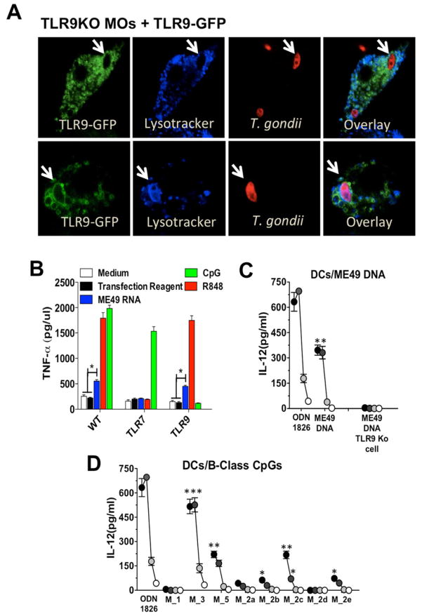
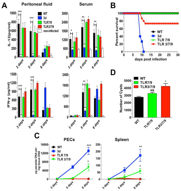
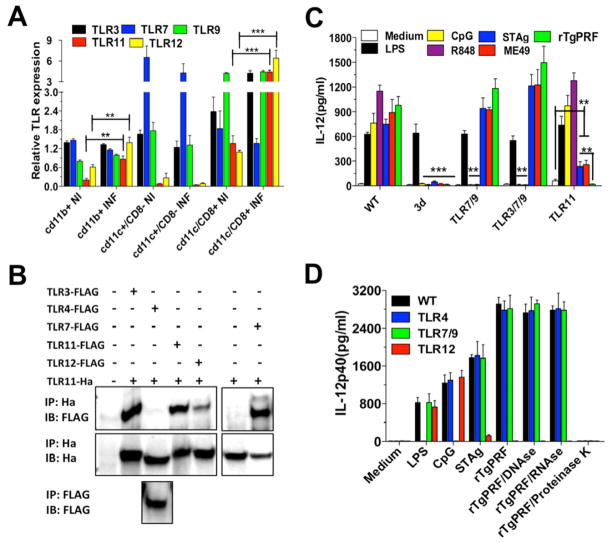
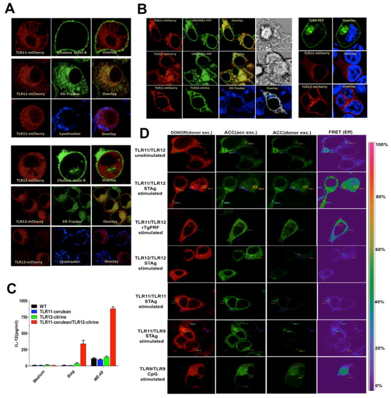
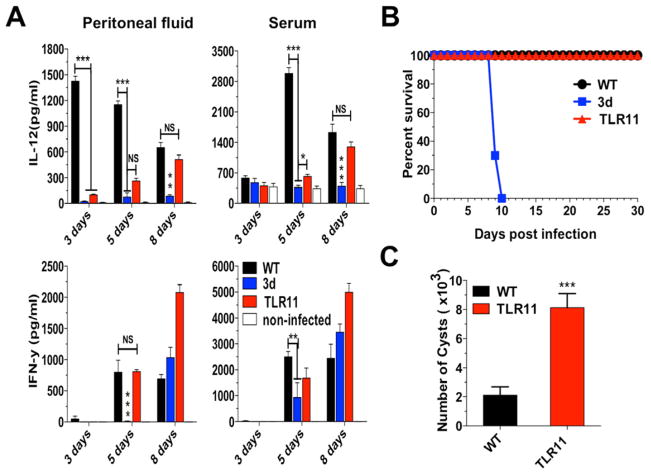
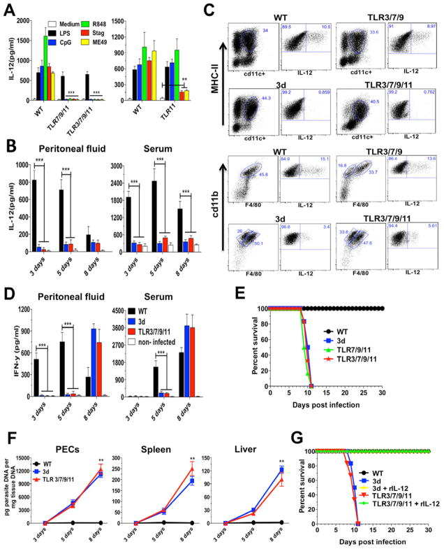
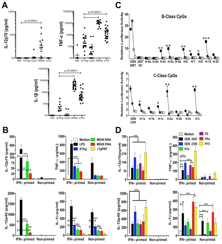
"Triple-defective" (3d) mice carrying a mutation in UNC93B1, a chaperone for the endosomal nucleic acid-sensing (NAS) Toll-like receptors TLR3, TLR7, and TLR9, are highly susceptible to Toxoplasma gondii infection. However, none of the single or even the triple NAS-TLR-deficient animals recapitulated the 3d susceptible phenotype to experimental toxoplasmosis. Investigating this further, we found that while parasite RNA and DNA activate innate immune responses via TLR7 and TLR9, TLR11 and TLR12 working as heterodimers are required for sensing and responding to Toxoplasma profilin. Consequently, the triple TLR7/TLR9/TLR11-deficient mice are highly susceptible to T. gondii infection, recapitulating the phenotype of 3d mice. Humans lack functional TLR11 and TLR12 genes. Consistently, human cells produce high levels of proinflammatory cytokines in response to parasite-derived RNA and DNA, but not to Toxoplasma profilin, supporting a more critical role for NAS-TLRs in human toxoplasmosis.


Figures

Figure 1
Figure 1. T. gondii RNA and DNA activate host cells via TLRs
(A) Confocal microscopy of immortalized TLR9−/− macrophages stably expressing TLR9-GFP and infected with CMTPX-stained T. gondii. Acidic compartments were stained with LysoTracker White-Blue. Arrows indicate internalized parasites. (B) Immortalized WT, TLR7 KO and TLR9 KO macrophages were stimulated with T.gondii RNA at 2 μg/ml complexed with DOTAP (Roche). (C) DCs were stimulated with T. gondii DNA complexed with DOTAP at 10, 5, 1 and 0.1 μg/ml. (D) DCs were stimulated with T. gondii derived oligonucleotides containing B-class mouse-like stimulatory CpG motifs at 3 mM (black circles), 1 mM (dark grey), 0.3 mM (light grey) and 0.1 mM (white circles). Cytokine levels were measured in the tissue culture supernatants at 24hrs after stimulation. (B-D) Data are represented as mean ± SD of three independent experiments. (*0.01 < p < 0.05, **0.001 < p < 0.01, ***p < 0.001). See also Supplementary Table1.
Figure 2
Figure 2. Mice deficient in TLR3/TLR7/TLR9 are only partially susceptible to T. gondii infection
Mice were infected intraperitoneally with 25 cysts of T. gondii ME49 strain. (A) Levels of IL-12p40 and IFNγ were measured in the peritoneal cavity exudate and sera at different times post-infection. (B) Combined survival data from WT (n=20), 3d (n=16) TLR7/9 (n=20) and TLR3/7/9 (n=20) mice from 4 independent experiments. (C) Quantitative real-time PCR analysis was performed on the indicated tissues collected from animals infected with T. gondii. Data are mean of three independent experiments. (D) Cysts counts in the brain determined at 30 days post-infection are the mean from four experiments. (A–D) Data are represented as mean ± SD. Asterisks indicate that difference is statistically significant, when comparing different mouse lineages infected with T. gondii, (NS: not significant, *0.01 < p < 0.05, **0.001 < p < 0.01, ***p < 0.001).
Figure 3
Figure 3. Endosomal TLRs are highly expressed in CD8α + DCs and upregulated upon T. gondii infection
(A) Real-time PCR was performed to determine the relative levels of TLR3, TLR7, TLR9, TLR11 and TLR12 mRNA expressed by CD11b+, CD11c+/CD8α+ and CD11c+/CD8α cells sorted from splenocytes from uninfected controls as well as infected (5 days post-infection) WT mice. TLR mRNA levels were normalized to Glyceraldehyde 3-phosphate dehydrogenase (GAPDH) mRNA. Data are represented as mean ±SD of three experiments. (B) HEK 293T cells were transfected with different pairs of plasmids, total lysates immunoprecipitated (IP) with anti-hemagglutinin (anti-HA, top) or anti-Flag (bottom), and analyzed by immunoblot (IB) with anti-Flag (top and bottom). The top membrane was then stripped and re-probed with anti-HA to ensure expression of hemagglutinin-tagged TLR11 (middle). (C) CD11c+ cells were purified from spleen of WT, 3d, TLR7/TLR9, TLR3/TLR7/TLR9 and TLR11 KO mice and stimulated with LPS (100 ng/ml) ODN CpG 1826 (1 μm), R848 (2 μm) or infected with ME49 tachyzoites (MOI 3:1). (D) CD11c+ cells purified from WT, TLR4, TLR7/TLR9 and TLR12 KO mice were stimulated with LPS (100 ng/ml), ODN CpG 1826 (1 μm), STAg (10 μg/ml), or rTgPRF (10 ng/ml) left untreated, or treated with DNAse (100 U/ml), RNAse (10 μg/ml) or Proteinase K (10 μg/ml). IL-12 levels were measured in the supernatant at 24 h after stimulation. (C–D) Data are represented as mean ± SD of four experiments. Asterisks indicate that difference is statistically significant, when comparing cytokines levels from WT to different KO mice, infected or not infected with T. gondii (*0.01 < p < 0.05, **0.001 < p < 0.01, ***p < 0.001). See also Figure S1.
Figure 4
Figure 4. Co-localization and heterodimerization of TLR11 and TLR12
(A) Immortalized macrophages were stably transfected with TLR11- or TLR12-mcherry and imaged post-staining with Cholera toxin B subunit FITC conjugate, LysoTracker White-Blue or ER-Tracker White-Blue. (B) HEK 293T cells transfected with TLR11-mCherry or TLR12-mcherry with UNC93B1-YFP or TLR11-mcherry and TLR12-citrine (left panel) or with TLR4-YFP and stained with Hoechst 33342 as nuclear marker (right panel). (C) Immortalized macrophages stably expressing either TLR11, TLR12 or both were stimulated in vitro with STAg (10 μg/ml) or exposed to live tachyzoites (MOI 3:1) of the ME49 strain and levels of IL-12p40 measured in the supernatants at 24 h post-stimulation. Data are represented as mean ± SD of three experiments. (D) HEK 293T cells were transfected with the plasmids encoding the proteins indicated in the figure; 48 h after transfection, cells were left unstimulated or were stimulated with STAg (10 μg/ml), rTgPRF (100 ng/ml) or CpG 1826 (1 μM) for TLR9/TLR9. FRET between the respective proteins was calculated by measuring sensitized emission (SE) fluorescence using the FRET SE wizard on the Leica SP2 confocal laser-scanning microscope. For each plasmid combination, cerulean (represented in red) was used as donor and citrine (represented in green) as acceptor. Data are from one representative experiment of four.
Figure 5
Figure 5. TLR11 mice are resistant to T. gondii infection
Mice were infected intraperitoneally with 25 cysts ME49 strain of T. gondii. (A) Levels of IL-12p40 and IFNγ were measured in the peritoneal cavity exudate and sera from uninfected as well as infected mice. (B) Combined survival data from WT (n=15), 3d (n=12) and TLR11 (n=15) mice from three independent experiments. (C) The cyst numbers in the brain were counted at 30 days post-infection and presented as mean from the three experiments. (A–C) Data are represented as mean ± SD. Asterisks indicate that difference is statistically significant, when comparing to unstimulated controls (NS: not significant, *0.01 < p < 0.05, **0.001 < p < 0.01, and ***p < 0.001).
Figure 6
Figure 6. Quadruple deficient mice have an impaired IL-12 and early IFNγ production and are highly susceptible to T. gondii infection
(A) CD11c+ cells were purified from spleen of WT, TLR7/TLR9/TLR11, TLR3/TLR7/TLR9 and TLR11 KO mice and stimulated with LPS (100 ng/ml), ODN CpG 1826 (1 μm), R848 (2 μm), STAg (10 μg/ml) or infected with ME49 tachyzoites (MOI 3:1). Data are represented as mean ± SD of two experiments. (B) Levels of IL-12p40 were measured in the peritoneal cavity exudate and sera from uninfected controls and infected mice. Data represent as mean ± SD of four experiments. (C) Mice infected with T. gondii were sacrificed at 5 days post-infection and peritoneal cells analyzed for IL-12 cellular source by intracellular cytokine staining. Dendritic cells (top) were gated for CD11c+/MHC-II+. Inflammatory monocytes (bottom) were gated first for GR1+ and then for CD11b+/F4/80+. Both populations were stained for IL-12p70. Data are from one representative experiment of three that yielded similar results. (D) Levels of IFNγ present in the peritoneal cavity exudate and sera from uninfected controls and infected mice. Data are represented as mean ± SD of four experiments. (E) Combined survival data from WT (n=15), 3d (n=10), TLR7/9/11 KO (n=10) and TLR3/7/9/11 KO (n=15) mice from at least two independent experiments. (F) Quantitative real-time PCR analysis was performed on the indicated tissues collected from animals infected with T. gondii. Data are represented as mean ± SD of three independent experiments. (G) WT, 3d and TLR3/TLR7/TLR9/TLR11 KO were infected i.p. with the ME49 strain of T. gondii (n=5 per group). Mice were treated with 100 ng of recombinant IL-12p70 or vehicle for 6 consecutive days, and mortality was evaluated. Data are from one representative experiment of two that yielded identical results. Asterisks indicate that difference is statistically significant, when comparing different mouse lineages infected with T. gondii. (**0.001 < p < 0.01, and ***p < 0.001). See also Figure S2.
Figure 7
Figure 7. DNA and RNA from T. gondii activate Human Peripheral Blood Mononuclear Cells (PBMCs)
(A) PBMCs purified from blood of clinically healthy donors were stimulated in vitro with STAg (10 μg/ml), CpG ODN 2007 (1 μM), R848 (2 μM) or LPS (100 ng/ml). Data are represented as mean ± SD. (B) PBMCs primed or not with recombinant human IFNγ (200 U/ml) were stimulated with LPS (100 ng/ml), STAg (10 μg/ml), ME49 RNA (2 μg/ml), ME49 DNA (5 μg/ml), or rTgPRF (1 μg/ml). Parasite RNA and DNA were complexed with DOTAP (Roche). (C) HEK 293T cells were stimulated with T. gondii derived oligonucleotides containing B or C-class human-like stimulatory CpG motifs at 3 μM (black circles), 1 mM (dark grey), 0.3 mM (light grey) and 0.1 mM (white circles). Asterisks indicate that differences are statistically significant when comparing stimulated cells with negative control - ODN 2007GC (top panel) or unstimulated cells (bottom panel). (D) PBMCs primed or not with recombinant human IFNγ (200 U/ml) were stimulated in vitro with T. gondii derived oligonucleotides containing B and C-class human-like stimulatory CpG motifs (5 mM). Cytokine levels were measured in the supernatants at 18 h after stimulation. (B-D) Data are represented as mean ± SD of three independent experiments (*0.01 < p < 0.05, **0.001 < p < 0.01, and ***p < 0.001). See also Supplementary Table 2.

References

    1. Alexopoulou L, Holt AC, Medzhitov R, Flavell RA. Recognition of double-stranded RNA and activation of NF-kappaB by Toll-like receptor 3. Nature. 2001;413:732–738. - PubMed
    1. Aosai F, Rodriguez Pena MS, Mun HS, Fang H, Mitsunaga T, Norose K, Kang HK, Bae YS, Yano A. Toxoplasma gondii-derived heat shock protein 70 stimulates maturation of murine bone marrow-derived dendritic cells via Toll-like receptor 4. Cell Stress Chaperones. 2006;11:13–22. - PMC - PubMed
    1. Bartholomeu DC, Ropert C, Melo MB, Parroche P, Junqueira CF, Teixeira SM, Sirois C, Kasperkovitz P, Knetter CF, Lien E, et al. Recruitment and endo-lysosomal activation of TLR9 in dendritic cells infected with Trypanosoma cruzi. J Immunol. 2008;181:1333–1344. - PubMed
    1. Benson A, Pifer R, Behrendt CL, Hooper LV, Yarovinsky F. Gut commensal bacteria direct a protective immune response against Toxoplasma gondii. Cell Host Microbe. 2009;6:187–196. - PMC - PubMed
    1. Brinkmann MM, Spooner E, Hoebe K, Beutler B, Ploegh HL, Kim YM. The interaction between the ER membrane protein UNC93B and TLR3, 7, and 9 is crucial for TLR signaling. J Cell Biol. 2007;177:265–275. - PMC - PubMed

Publication types

MeSH terms

LinkOut - more resources