. 2013 May;10(5):728-37.
doi: 10.1016/j.hrthm.2012.12.033. Epub 2013 Jan 2.

Dysfunctional potassium channel subunit interaction as a novel mechanism of long QT syndrome

Affiliations

Dysfunctional potassium channel subunit interaction as a novel mechanism of long QT syndrome

Michael Hoosien et al. Heart Rhythm. 2013 May.

Abstract

Background: The slowly-activating delayed rectifier current IKs contributes to repolarization of the cardiac action potential, and is composed of a pore-forming α-subunit, KCNQ1, and a modulatory β-subunit, KCNE1. Mutations in either subunit can cause long QT syndrome, a potentially fatal arrhythmic disorder. How KCNE1 exerts its extensive control over the kinetics of IKs remains unresolved

Objective: To evaluate the impact of a novel KCNQ1 mutation on IKs channel gating and kinetics

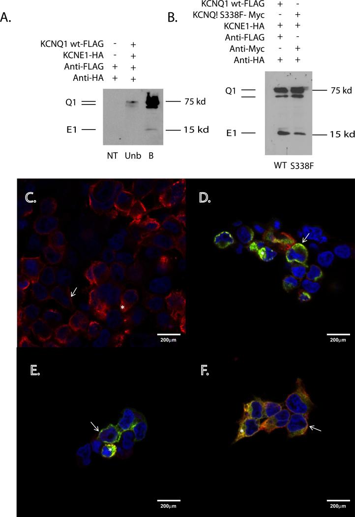
Methods: KCNQ1 mutations were expressed in Xenopus oocytes in the presence and absence of KCNE1. Voltage clamping and MODELLER software were used to characterize and model channel function. Mutant and wt genes were cloned into FLAG, Myc and HA expression vectors to achieve differential epitope tagging, and expressed in HEK293 cells for immunohistochemical localization and surface biotinylation assay.

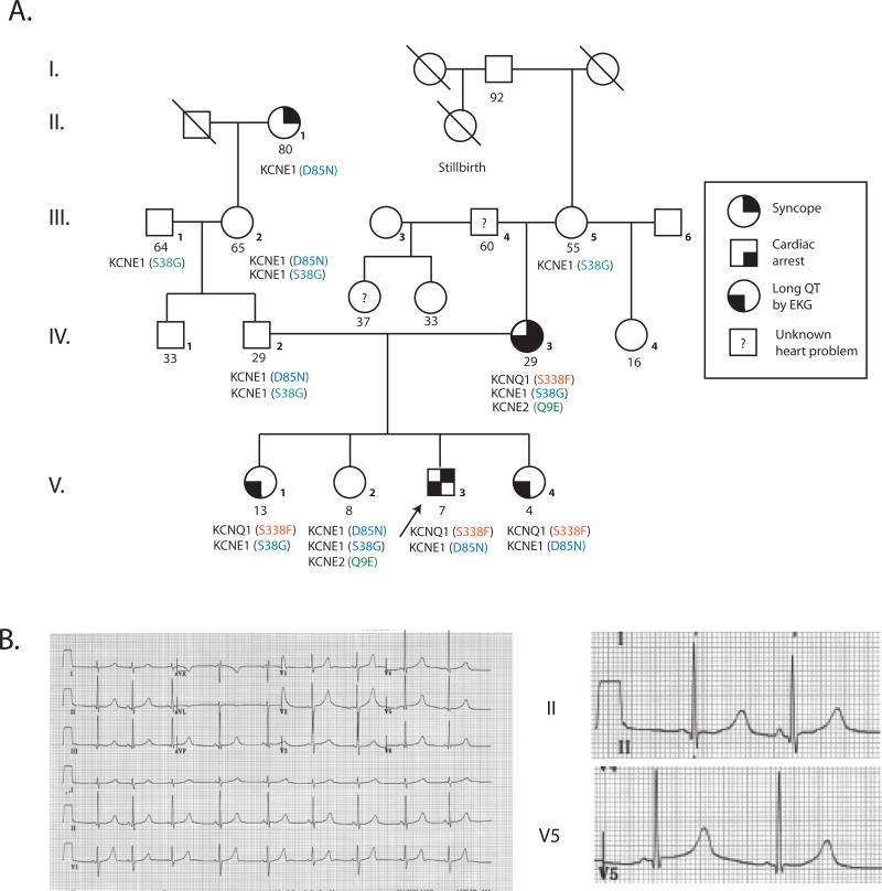
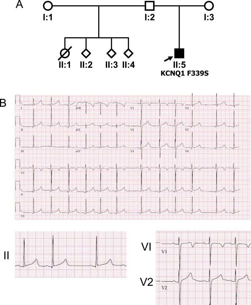
Results: We identified 2 adjacent mutations, S338F and F339S, in the KCNQ1 S6 domain in unrelated probands. The novel KCNQ1 S338F mutation segregated with prolonged QT interval and torsade de pointes; the second variant, F339S, was associated with fetal bradycardia and prolonged QT interval, but no other clinical events. S338F channels expressed in Xenopus oocytes had slightly increased peak conductance relative to wild type, with a more positive activation voltage. F339S channels conducted minimal current. Unexpectedly, S338F currents were abolished by co-expression with intact WT KCNE1 or its C-terminus (aa63-129), despite normal membrane trafficking and surface co-localization of KCNQ1 S338F and wt KCNE1. Structural modeling indicated that the S338F mutation specifically alters the interaction between the S6 domain of one KCNQ1 subunit and the S4-S5 linker of another, inhibiting voltage-induced movement synergistically with KCNE1 binding.

Conclusions: A novel KCNQ1 mutation specifically impaired channel function in the presence of KCNE1. Our structural model shows that this mutation effectively immobilizes voltage gating by an inhibitory interaction that is additive with that of KCNE1. Our findings illuminate a previously unreported mechanism for LQTS, and validate recent theoretical models of the closed state of the KCNQ1:KCNE1 complex.


Figures

Figure 1
Figure 1. Proband 1 pedigree and ECG
A. Pedigree B. 12-lead electrocardiogram from proband 1 at 7 years of age.
Figure 2
Figure 2. Proband 2 Pedigree and ECG
A. Pedigree B. 12-lead electrocardiogram from proband 2.
Figure 3
Figure 3. Adjacent novel mutations in the KCNQ1 S6 transmembrane domain
A. Partial KCNQ1 exon 7 DNA sequence from proband 1. B. Partial KCNQ1 exon 7 DNA sequence from proband 2.
Figure 4
Figure 4. KCNQ1 S338F mutation induces a dysfunctional interaction with KCNE1
A. Homomeric currents generated by WT KCNQ1 and S338F and F339S variants. B. Addition of KCNE1 activates WT and suppresses mutant KCNQ1 currents. C-E. Current density–voltage relationships for KCNQ1 and IKs currents. F. KCNE1 C-terminal domain represses KCNQ1 S338F currents.
Figure 5
Figure 5. Dominant negative effects of KCNQ1 S338F and F339s mutations
A. Current density-voltage relationships for WT, mutant and mixed α-subunits. B. Effect of KCNE1 co-expression on WT and mutant KCNQ1 current density. C. Inhibitory effect of KCNQ1 mutants on KCNE1-generated currents. D. Dominant negative effect of S338F on WT KCNQ1/IKs current. E. Differential effect of KCNE1 D85N variant on WT and S338F mutant KCNQ1/IKs currents.
Figure 6
Figure 6. KCNQ1 S338F-KCNE1 channels traffic normally to plasma membrane
A. Biotinylation identifies membrane-localized WT KCNQ1-KCNE1 channels. NT = nontransfected, WT = KCNQ1 WT-FLAG, Unb = unbound and B = bound fraction of cell lysates. B. Equivalent membrane localization of WT and S338F mutant KCNQ1 in the presence of KCNE1. (Left lane) Biotinylated fraction of WT KCNQ1-FLAG and KCNE1-HA-transfected HEK293 cells. (Right lane) Biotinylated fraction of KCNQ1- S338F-MYC and KCNE1-HA-transfected HEK293 cells. C-F. Membrane co-localization of WT and S338F mutant KCNQ1 in the presence of KCNE1. C. Membrane localization of WT KCNQ1-FLAG in the presence of WT KCNE1-HA. Arrows= Fluorescent signal at the cell surface. Star = signal within the intracellular compartment. The nucleus has been counterstained with DAPI (blue). D. Membrane co-localization of KCNQ1 S338F and KCNE1. Green = S338F KCNQ1-MYC; red = WT KCNE1-HA. Co-localization (yellow) can be seen at the cell surface (arrow) and within the interior of the cell (star). E. Membrane co-localization of KCNQ1 S338F and KCNE1 in the presence of WT KCNQ1. Red = anti-HA (KCNE1). Green = anti-MYC (S338F KCNQ1). F. Membrane co-localization of KCNQ1 S338F and KCNQ1 WT in the presence of KCNE1. Triple-transfected cells as in E were imaged for S338F (MYC, green) and WT KCNQ1 (FLAG, red). Again note signal co-localization at the cell membrane (arrow) and to a lesser extent within the cell interior (star).
Figure 7
Figure 7. Structural modeling of KCNQ1 mutant channels. A and B. Model of WT KCNQ1 channel in the closed state
A. Bottom view, as visualized along an axis perpendicular to the plane of the cell membrane from the cytosolic side. Dashed circles and connecting arrows show direction of rotation of the VS domain during channel opening. Red dot denotes the central pore. B. Side view, in the plane of the cell membrane. Blue, green, cyan and purple: individual monomers. Gray: the S4-S5 helical linker connecting the VS domain (helices S1-S4) to the pore forming domain (helices S5/P/S6) within each monomer. Red arrow: path of potassium ions. C and D. Impact of the S338F variant on KCNQ1 channel structure. In C and D, the interface of two monomers (cyan and green) is highlighted within the tetrameric KCNQ1. Grey: S4-S5 helical linker. C. Model of KCNQ1 WT in the closed state. D. Model of KCNQ1 S338F in the closed state. Red: sidechain moieties of residues S338/F338 in the S6 helix are shown interacting through van der Waals forces with residues W248/L251 residues in the S4-S5 helical linker of an adjacent monomer. E and F. Impact of the F339S variant on KCNQ1 channel structure. In these models, only one monomer is shown (blue) so as to highlight inter-helical interactions within each monomer. Gray: S4-S5 helical linker. Red: sidechain moieties of F339/S339 and T265/I268 involved in intramolecular van der Waals contacts between S5 and S6 helices. E. Model of KCNQE1 WT in the closed state. F. Model of KCNQ1 F339S in the closed state.

Comment in

References

    1. Modell SM, Lehmann MH. The long QT syndrome family of cardiac ion channelopathies: a HuGE review. Genet Med. 2006;8:143–155. - PubMed
    1. Moss AJ, Shimizu W, Wilde AA, et al. Clinical aspects of type-1 long-QT syndrome by location, coding type, and biophysical function of mutations involving the KCNQ1 gene. Circulation. 2007;115:2481–2489. - PMC - PubMed
    1. Sanguinetti MC, Curran ME, Zou A, et al. Coassembly of K(V)LQT1 and minK (IsK) proteins to form cardiac I(Ks) potassium channel. Nature. 1996;384:80–83. - PubMed
    1. Barhanin J, Lesage F, Guillemare E, Fink M, Lazdunski M, Romey G. K(V)LQT1 and lsK (minK) proteins associate to form the I(Ks) cardiac potassium current. Nature. 1996;384:78–80. - PubMed
    1. Nerbonne JM, Kass RS. Molecular physiology of cardiac repolarization. Physiol Rev. 2005;85:1205–1253. - PubMed

Publication types

Substances

LinkOut - more resources